. 2014;10(3):734-9.
doi: 10.4161/hv.27316. Epub 2013 Dec 3.

Characterization of virus-like particles in GARDASIL® by cryo transmission electron microscopy

Affiliations

Characterization of virus-like particles in GARDASIL® by cryo transmission electron microscopy

Qinjian Zhao et al. Hum Vaccin Immunother. 2014.

Abstract

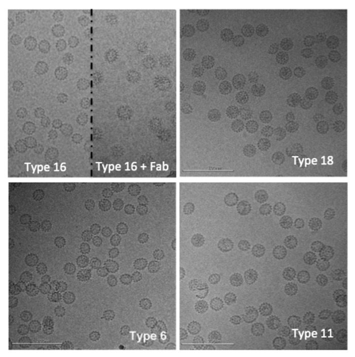
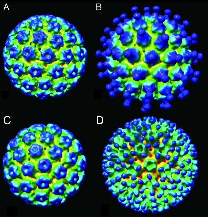
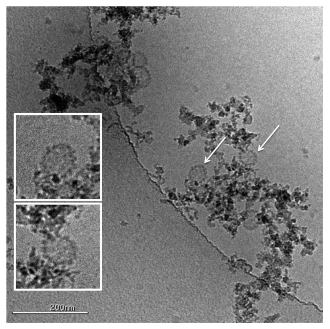
Cryo-transmission electron microscopy (cryoTEM) is a powerful characterization method for assessing the structural properties of biopharmaceutical nanoparticles, including Virus Like Particle-based vaccines. We demonstrate the method using the Human Papilloma Virus (HPV) VLPs in GARDASIL®. CryoTEM, coupled to automated data collection and analysis, was used to acquire images of the particles in their hydrated state, determine their morphological characteristics, and confirm the integrity of the particles when absorbed to aluminum adjuvant. In addition, we determined the three-dimensional structure of the VLPs, both alone and when interacting with neutralizing antibodies. Two modes of binding of two different neutralizing antibodies were apparent; for HPV type 11 saturated with H11.B2, 72 potential Fab binding sites were observed at the center of each capsomer, whereas for HPV 16 interacting with H16.V5, it appears that 60 pentamers (each neighboring 6 other pentamers) bind five Fabs per pentamer, for the total of 300 potential Fab binding sites per VLP.

Keywords: CryoTEM; Gardasil; VLP; adjuvant; aluminum; epitope; structure.


Figures

None
Figure 1. Representative cryo-electron microscopy images of human papillomavirus virus-like particles (L1 types 16, 18, 6, and 11). In the top left panel, the image is split to show type 16 particles on the left and type 16 particles decorated with an antibody fragment on the right. Scale bar is 200 nm.
None
Figure 2. Three-dimensional reconstructions of (A) HPV11 used 5135 particle images in the reconstruction and the resolution based on the FSC0.5 criteria was 1.3 nm and the EMDB deposition number is 28367; (B) HPV11 decorated with antibody fragment H11.B2 B2 (6009 particles, FSC0.5 = 2.0 nm, EMDB 28368); (C) HPV16 (5135 particles, FSC0.5 = 1.3 nm, EMDB 28369) (d) HPV16 decorated with antibody fragment H16.V5 (6009 particles, FSC0.5 = 2.0 nm, EMDB 28370).
None
Figure 3. HPV VLPs can be observed directly interacting with the alum adjuvant. The VLPs indicated by the arrows are shown at larger scale in the insets indicating that the VLPs are intact and retain their morphology and spherical shape upon absorption onto the adjuvant.

References

    1. Dunne EF, Datta SD, E Markowitz L. A review of prophylactic human papillomavirus vaccines: recommendations and monitoring in the US. Cancer. 2008;113(Suppl):2995–3003. doi: 10.1002/cncr.23763. - DOI - PubMed
    1. Kirnbauer R, Booy F, Cheng N, Lowy DR, Schiller JT. Papillomavirus L1 major capsid protein self-assembles into virus-like particles that are highly immunogenic. Proc Natl Acad Sci U S A. 1992;89:12180–4. doi: 10.1073/pnas.89.24.12180. - DOI - PMC - PubMed
    1. Deschuyteneer M, Elouahabi A, Plainchamp D, Plisnier M, Soete D, Corazza Y, Lockman L, Giannini S, Deschamps M. Molecular and structural characterization of the L1 virus-like particles that are used as vaccine antigens in Cervarix™, the AS04-adjuvanted HPV-16 and -18 cervical cancer vaccine. Hum Vaccin. 2010;6:407–19. doi: 10.4161/hv.6.5.11023. - DOI - PubMed
    1. McCarthy MP, White WI, Palmer-Hill F, Koenig S, Suzich JA. Quantitative disassembly and reassembly of human papillomavirus type 11 viruslike particles in vitro. J Virol. 1998;72:32–41. - PMC - PubMed
    1. Finch JT, Klug A. The structure of viruses of the papilloma-polyoma type 3. Structure of rabbit papilloma virus, with an appendix on the topography of contrast in negative-staining for electron-microscopy. J Mol Biol. 1965;13:1–12. doi: 10.1016/S0022-2836(65)80075-4. - DOI - PubMed

Publication types

MeSH terms

Substances