. 2014 Jan 17;289(3):1866-75.
doi: 10.1074/jbc.M113.505578. Epub 2013 Dec 3.

Cross-talk between aryl hydrocarbon receptor and the inflammatory response: a role for nuclear factor-κB

Affiliations

Cross-talk between aryl hydrocarbon receptor and the inflammatory response: a role for nuclear factor-κB

Christoph F A Vogel et al. J Biol Chem. .

Abstract

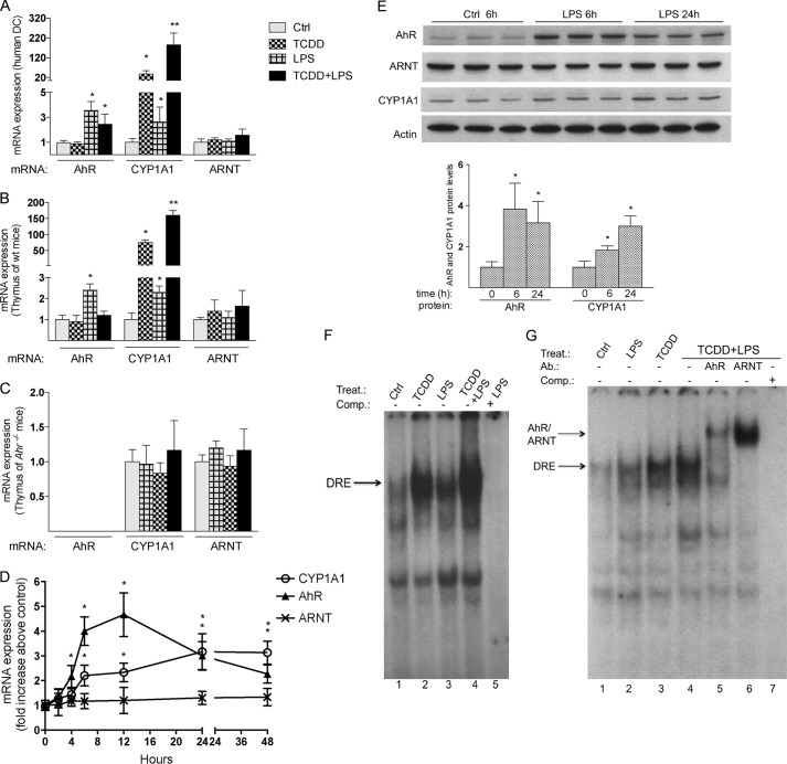
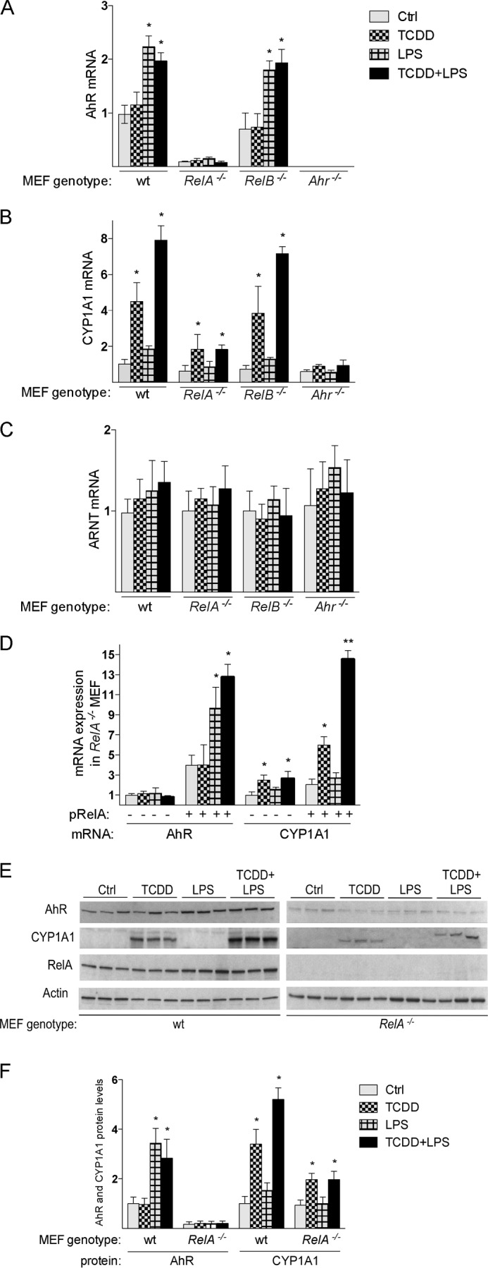
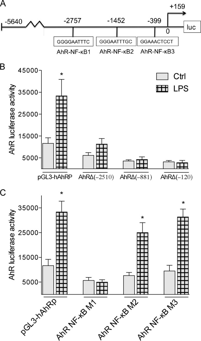
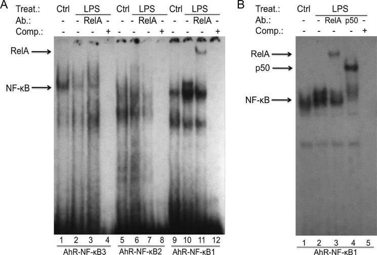
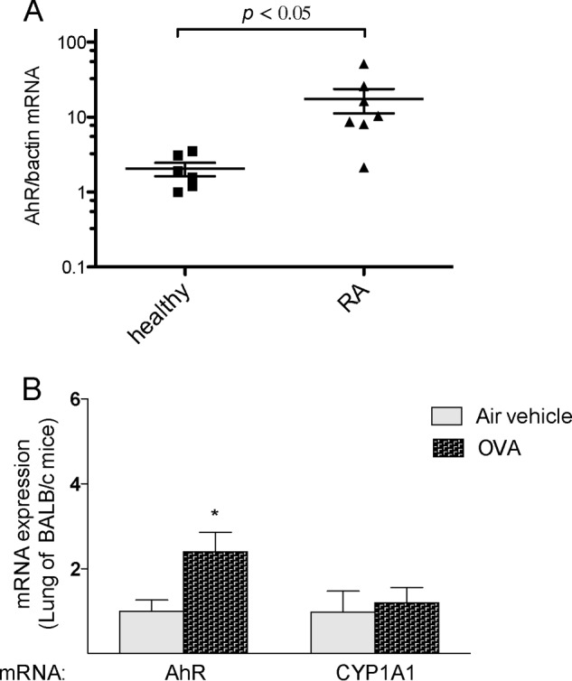
The aryl hydrocarbon receptor (AhR) is involved in the regulation of immune responses, T-cell differentiation, and immunity. Here, we show that inflammatory stimuli such as LPS induce the expression of AhR in human dendritic cells (DC) associated with an AhR-dependent increase of CYP1A1 (cytochrome P4501A1). In vivo data confirmed the elevated expression of AhR by LPS and the LPS-enhanced 2,3,7,8-tetrachlorodibenzo-p-dioxin (TCDD)-mediated induction of CYP1A1 in thymus of B6 mice. Inhibition of nuclear factor-κB (NF-κB) repressed both normal and LPS-enhanced, TCDD-inducible, AhR-dependent gene expression and canonical pathway control of RelA-regulated AhR-responsive gene expression. LPS-mediated induction of AhR was NF-κB-dependent, as shown in mouse embryonic fibroblasts (MEFs) derived from Rel null mice. AhR expression and TCDD-mediated induction of CYP1A1 was significantly reduced in RelA-deficient MEF compared with wild type MEF cells and ectopic expression of RelA restored the expression of AhR and induction of CYP1A1 in MEF RelA null cells. Promoter analysis of the human AhR gene identified three putative NF-κB-binding elements upstream of the transcription start site. Mutation analysis of the AhR promoter identified one NF-κB site as responsible for mediating the induction of AhR expression by LPS and electrophoretic shift assays demonstrated that this NF-κB motif is recognized by the RelA/p50 heterodimer. Our results show for the first time that NF-κB RelA is a critical component regulating the expression of AhR and the induction of AhR-dependent gene expression in immune cells illustrating the interaction of AhR and NF-κB signaling.

Keywords: Aryl Hydrocarbon Receptor; Dendritic Cells; Gene Regulation; Inflammation; NF-Kappa B (NF-KB).


Figures

FIGURE 1.
FIGURE 1.
Increased expression of AhR and CYP1A1 by LPS in human DC and thymus of B6 mice. A, LPS induces AhR and CYP1A1 in human DC. Human monocyte-derived DC were generated as described under “Experimental Procedures.” DC were treated with 10 nm TCDD for 7 days or 0.05 μg/ml LPS for 24 h. TCDD-treated DC were co-treated with LPS for 24 h. B6 wild type (B) and Ahr−/− (C) mice were treated with 15 μg/kg TCDD for 24 h or with 0.5 mg/kg LPS for 6 h. For co-treatment, mice were treated with 15 μg/kg TCDD for 18 h and then treated with 0.5 mg/kg LPS for 6 h. D, time course study of AhR, ARNT, and CYP1A1 mRNA induction in U937-derived DC. Cells were treated for 1 to 48 h with 0.1 μg/ml LPS or 1 μl/ml water (Control). E, Western blot analysis of AhR, ARNT, and CYP1A1 protein level in human DC. 25 μg of whole cell protein was loaded per lane. AhR and CYP1A1 protein levels were quantitated and normalized to actin. Values represent the mean ± S.D. of three independent experiments. An asterisk indicates significantly different from control cells (p < 0.05). Double asterisks indicate significantly higher than only TCDD-treated cells (p < 0.05). F, LPS-induced AhR binding to a DRE consensus element of the Cyp1a1 promoter. Nuclear extracts from untreated control (lane 1) and LPS-treated (lanes 3 and 5) U937-derived DC were used for EMSA. Cells were treated with 1 nm TCDD for 1 h or with 0.1 μg/ml LPS for 6 h. A possible enhancement of TCDD-induced AhR-binding activity as shown in lane 2 was tested by treatment of cells with LPS (0.1 μg/ml) for 6 h followed by treatment with TCDD (1 nm) for 1 h (lane 4). EMSA was performed using double-stranded, 32P-labeled oligonucleotide containing the DRE binding sequence of the rat Cyp1a1 promoter. To confirm specificity, a 100-fold excess of unlabeled DRE oligonucleotide (lane 5) was added. G, the specific binding of AhR and ARNT was identified by EMSA supershift analyses using AhR- and ARNT-specific antibodies (lanes 5 and 6). For EMSA, one representative experiment of three independently performed experiments is shown. Ab., antibody; Comp., competition; Ctrl, control; Treat., treatment.
FIGURE 2.
FIGURE 2.
NF-κB RelA dependent expression of AhR. A, U937-derived DC were treated with 0.1 μg/ml LPS, 1 nm TCDD, and co-treated with TCDD and LPS for 6 h in absence or presence of 200 μm PDTC. PDTC or water was added 30 min prior to the addition of DMSO as vehicle, TCDD, or LPS. AhR, ARNT, and CYP1A1 mRNA levels were analyzed using real-time PCR. B, effect of NF-κB inhibitors on TCDD-induced and LPS-enhanced DRE luciferase reporter activity. U937-derived DC were incubated with carrier solvent alone (1 μl/ml), 0.1 μg/ml LPS, 1 nm TCDD, 200 μm PDTC, 5 μg/ml CAPE, 50 μm CAPS, or the indicated combination. TCDD was dissolved in DMSO, LPS, and PDTC in water, and CAPE and CAPS were dissolved in ethanol. Cells were treated with LPS for 16 h and with TCDD for 4 h. PDTC, CAPE, and CAPS were added 30 min prior to LPS and TCDD. Luciferase activity was determined as described under “Experimental Procedures.” Values are expressed as relative luminescence units/mg protein and represent the mean ± S.D. of triplicate determinations. C, siRNA-mediated RelA gene ablation was performed in U937-derived DC. After transient transfection for 48 h, cells were treated with 1 nm TCDD, 0.1 μg/ml LPS, or 0.1% DMSO (Ctrl; control) for 6 h. mRNA levels of AhR and CYP1A1 were determined using real-time PCR. Values for AhR and CYP1A1 mRNA expression are normalized to the expression of β-actin. Values are the mean ± S.D. of three independent experiments. Single asterisk indicates significantly different from control (p < 0.05); double asterisks indicate significantly higher than only TCDD-treated cells (p < 0.05); triple asterisks indicate significantly lower than only TCDD- or LPS-treated cells (p < 0.05). D, RelA protein ablation was confirmed by Western blot analysis of U937-derived DC transfected with RelA siRNA (siRelA) or scrambled siRNA (siCtrl) for 48 h. 25 μg of whole cell protein was loaded per lane.
FIGURE 3.
FIGURE 3.
Expression of AhR and CYP1A1 in MEF cells from wt and Rel null mice. A, expression of AhR mRNA; B, CYP1A1 mRNA; C, ARNT mRNA in MEF cells were analyzed using real-time PCR. MEF cells derived from wild type (wt), RelA-deficient mice (RelA−/−), RelB-deficient mice (RelB−/−), and AhR-deficient mice (Ahr−/−) were treated with 1 nm TCDD, 0.1 μg/ml LPS, or 0.1% DMSO (Ctrl; control) for 6 h. D, induction of AhR and CYP1A1 is restored in MEF RelA−/− cells after transient transfection with a RelA. Cells were transiently transfected with a control or RelA expression plasmid (pRelA) for 24 h and treated with 1 nm TCDD, 0.1 μg/ml LPS, or 0.1% DMSO (Ctrl) for 6 h. E, Western blot analysis of AhR, CYP1A1, and RelA protein levels in WT and RelA−/− MEF. 25 μg of whole cell protein was loaded per lane. F, AhR and CYP1A1 protein levels were quantitated and normalized to actin. Values represent the mean ± S.D. of three independent experiments. A single asterisk indicates significantly different from control cells (p < 0.05). Values for AhR, ARNT, and CYP1A1 mRNA expression are normalized to the expression of β-actin. Values are the mean ± S.D. of three independent experiments. A single asterisk indicates significantly different from control cells (p < 0.05).
FIGURE 4.
FIGURE 4.
LPS-specific effects on deletion and mutation constructs of the human AhR promoter. A, schematic illustration of the full-length promoter construct of the human AhR gene containing 5640 bp upstream of the transcriptional start site (indicated by an arrow) cloned into a luciferase (luc) reporter vector. Positions of the three putative NF-κB recognition sites are presented. B, effect of LPS on AhR deletion constructs. U937-derived DC were transiently transfected with pGL3-hAhRP, and the deletion constructs AhRΔ(−2510), AhRΔ(−881), or AhRΔ(−120). C, effect of LPS on AhR promoter constructs mutated in NF-κB-binding sites. U937-derived DC were transiently transfected with equimolar amounts of the designated deletion or mutation constructs and treated with 100 ng/ml LPS for 6 h. Mean + S.D. of three independent experiments are given. A single asterisk indicates significantly different from control (p < 0.05).
FIGURE 5.
FIGURE 5.
LPS induces nuclear protein binding to a NF-κB-binding element of the AhR promoter. Nuclear extracts from untreated control (lanes 1, 5, and 9) and LPS-treated (lanes 2, 6, and 10) U937-derived DC were used for EMSA. A, EMSA was performed using double-stranded, 32P-labeled oligonucleotides containing the AhR-NF-κB1, AhR-NF-κB2, or AhR-NF-κB3 binding sequence of the human AhR promoter. A possible binding of RelA was identified by supershift analyses using RelA-specific antibodies (lanes 3, 7, and 11). B, EMSA was performed using double-stranded, 32P-labeled oligonucleotides containing the AhR-NF-κB1 site. The bands corresponding to the specific LPS-induced RelA and p50 NF-κB subunits are indicated by arrows (lanes 3 and 4). To confirm specificity, a 100-fold excess of unlabeled AhR-NF-κB oligonucleotides from the AhR promoter was added (lane 5). One representative experiment of three independently performed experiments is shown. Ab., antibody; Comp., competition; Ctrl, control; Treat., treatment.
FIGURE 6.
FIGURE 6.
Elevated expression of AhR in inflammatory disease. A, AhR expression in PBMCs from healthy and RA patients. Total RNA was harvested from PBMCs from six healthy and seven RA patients matched by gender and age. B, expression of AhR in lungs of mice after ovalbumin (OVA)-induced allergic lung inflammation and airway hyperreactivity. BALB/c mice (five mice per group) were sensitized by intraperitoneal (intraperitoneal) injection of ovalbumin and alum solution for 4 weeks and then exposed to ovalbumin aerosol six times over a period of 12 days as described elsewhere (26). Age-matched control animals were injected intraperitoneal with ovalbumin + alum adjuvant (sensitized) and were exposed only to filtered air. Experiments were performed in triplicate for each of the samples. AhR and CYP1A1 mRNA levels were analyzed by real-time PCR. A single asterisk indicates significantly different from control (p < 0.05).

References

    1. Schmidt J. V., Carver L. A., Bradfield C. A. (1993) Molecular characterization of the murine Ahr gene. Organization, promoter analysis, and chromosomal assignment. J. Biol. Chem. 268, 22203–22209 - PubMed
    1. Fitzgerald C. T., Nebert D. W., Puga A. (1998) Regulation of mouse Ah receptor (Ahr) gene basal expression by members of the Sp family of transcription factors. DNA Cell. Biol. 17, 811–822 - PubMed
    1. FitzGerald C. T., Fernandez-Salguero P., Gonzalez F. J., Nebert D. W., Puga A. (1996) Differential regulation of mouse Ah receptor gene expression in cell lines of different tissue origins. Arch. Biochem. Biophys. 333, 170–178 - PubMed
    1. Marshall N. B., Kerkvliet N. I. (2010) Dioxin and immune regulation: emerging role of aryl hydrocarbon receptor in the generation of regulatory T cells. Ann. N. Y. Acad. Sci. 1183, 25–37 - PMC - PubMed
    1. Ruby C. E., Leid M., Kerkvliet N. I. (2002) 2,3,7,8-Tetrachlorodibenzo-p-dioxin suppresses tumor necrosis factor-α and anti-CD40-induced activation of NF-κB/Rel in dendritic cells: p50 homodimer activation is not affected. Mol. Pharmacol. 62, 722–728 - PubMed

Publication types

MeSH terms