. 2013 Dec 17;110(51):20831-6.
doi: 10.1073/pnas.1307960110. Epub 2013 Dec 3.

Dysferlin stabilizes stress-induced Ca2+ signaling in the transverse tubule membrane

Affiliations

Dysferlin stabilizes stress-induced Ca2+ signaling in the transverse tubule membrane

Jaclyn P Kerr et al. Proc Natl Acad Sci U S A. .

Abstract

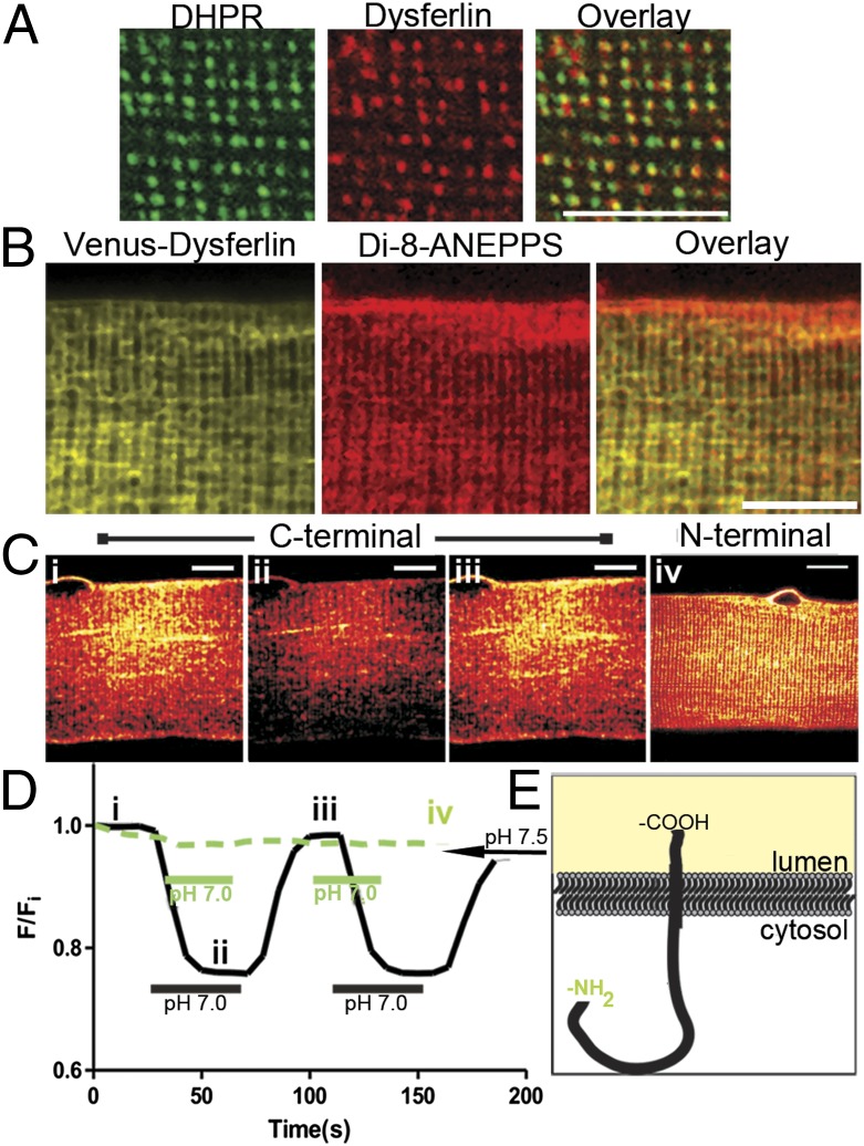
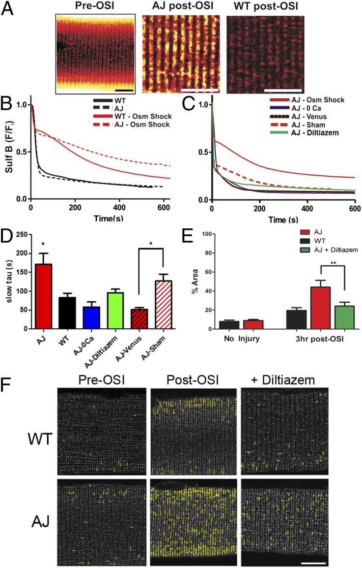
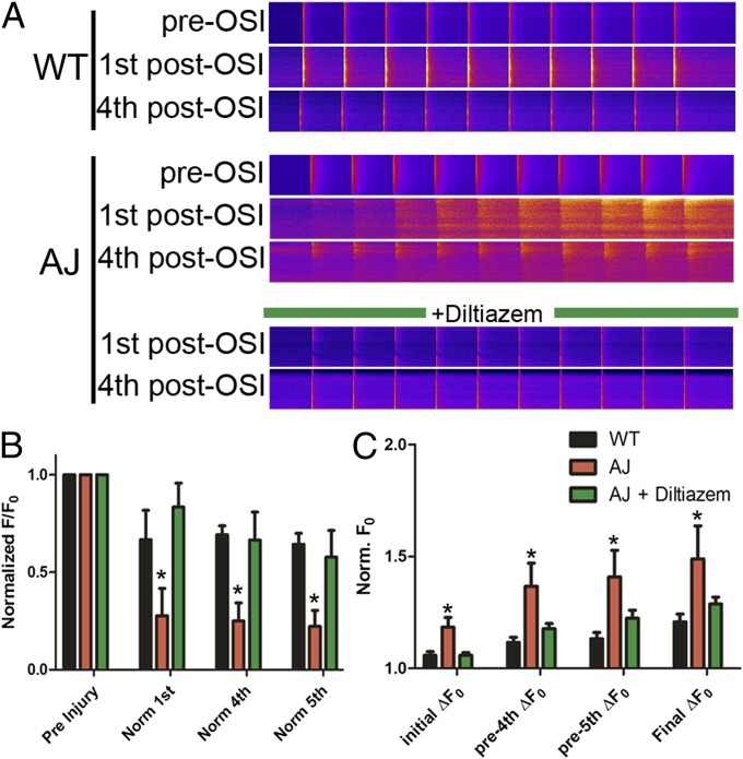
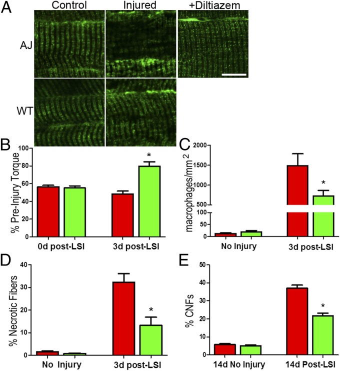
Dysferlinopathies, most commonly limb girdle muscular dystrophy 2B and Miyoshi myopathy, are degenerative myopathies caused by mutations in the DYSF gene encoding the protein dysferlin. Studies of dysferlin have focused on its role in the repair of the sarcolemma of skeletal muscle, but dysferlin's association with calcium (Ca(2+)) signaling proteins in the transverse (t-) tubules suggests additional roles. Here, we reveal that dysferlin is enriched in the t-tubule membrane of mature skeletal muscle fibers. Following experimental membrane stress in vitro, dysferlin-deficient muscle fibers undergo extensive functional and structural disruption of the t-tubules that is ameliorated by reducing external [Ca(2+)] or blocking L-type Ca(2+) channels with diltiazem. Furthermore, we demonstrate that diltiazem treatment of dysferlin-deficient mice significantly reduces eccentric contraction-induced t-tubule damage, inflammation, and necrosis, which resulted in a concomitant increase in postinjury functional recovery. Our discovery of dysferlin as a t-tubule protein that stabilizes stress-induced Ca(2+) signaling offers a therapeutic avenue for limb girdle muscular dystrophy 2B and Miyoshi myopathy patients.

Keywords: dihydropyridine receptor; excitation–contraction coupling; muscle injury; triad junction.


Conflict of interest statement

The authors declare no conflict of interest.

Figures

Fig. 1.
Fig. 1.
Dysferlin is a t-tubule protein. (A) Double-immunofluorescence labeling shows colabeling of dysferlin with DHPR. Analysis with Mander’s coefficient shows that ∼47% of DHPR colocalizes with dysferlin (M1 = 0.475 ± 0.016 SEM; n = 8). (B) FDB fibers were electroporated with cDNA plasmid encoding Venus-tagged dysferlin. Isolated fibers were treated with the lipophilic dye, di-8-ANEPPS. Venus-dysferlin colocalized with di-8-ANEPPS at the t-tubule doublets, but to a lesser extent at the sarcolemma. (C) FDB fibers overexpressing fusion proteins of pHluorin and dysferlin. Dysferlin with pHluorin at both the C terminus (i–iii) and N terminus (iv) show localization to doublets. (D) C-terminal pHluorin-dysferlin (black trace, i–iii in D) responds to changes in extracellular pH (∼25% decrease in fluorescence from pH 7.5 to pH 7.0); no significant change with the N-terminal pHluorin (green trace) was apparent. (E) A schematic representation of dysferlin shows the orientation of the protein in the t-tubule membrane. (Scale bars, 10 μm.)
Fig. 2.
Fig. 2.
Dysferlin-null myofibers are more susceptible to t-tubule damage in vitro. (A) SulfB revealed that the overall organization of the t-tubule does not differ significantly between AJ and WT (Left). Following OSI, a large amount of SulfB remains in the AJ t-tubules (Center) but not in WT (Right). (B) The kinetics of efflux of SulfB from t-tubules during washout (black lines) before OSI are indistinguishable between dysferlin-null and controls. Following osmotic shock, the mobility of SulfB was greatly reduced in AJ fibers (red dashed line) compared with WT controls (red solid line). (C) Dysferlin-null expressing Venus-dysferlin were resistant to osmotic shock (red line and black dashed line; AJ-Venus) compared with sham-transfected fibers (dashed red line; AJ-sham). In AJ fibers, removal of extracellular Ca2+ (blue line) or pretreatment (30 min) with diltiazem (green line) normalized dye clearance to near control rates. (D) Pooled data for the time constants (tau) of the slower phase of dye release under the different conditions assayed. (E and F) Dysferlin-null and WT FDB fibers immunolabeled for DHPR (grayscale; see also Fig. S2). The images were analyzed to assess areas of clustered DHPR (shown in yellow highlights) following OSI. Pooled data (E) show that only 10–15% of the area in uninjured dysferlin-null or control fibers is disrupted. Clustering of DHPR in dysferlin-null fibers increased to 44% at 3 h post-OSI. Treatment with diltiazem before and during osmotic shock decreased DHPR disruption in injured dysferlin-null fibers to levels indistinguishable from controls. We found no change in dysferlin distribution following OSI in WT FDB fibers (Fig. S2C). (Scale bars, 10 µm.) *P < 0.05, **P < 0.01.
Fig. 3.
Fig. 3.
Dysferlin-null myofibers exhibit altered Ca2+ regulation following injury. (A) Representative linescans of Ca2+ transients derived from WT (Top) and AJ (Middle) myofibers before OSI (“pre-OSI” rows), obtained with fibers loaded with Fluo-4, demonstrate normal Ca2+ homeostasis. Following OSI (“1st post-OSI” and “4th post-OSI” rows), both WT and AJ myofibers exhibit altered Ca2+ homeostasis, with the AJ myofibers demonstrating increased cytosolic Ca2+ and depressed transients. Diltiazem treatment (Bottom) largely prevents these changes in the AJ myofibers following OSI, resulting in transients that closely resemble WT. (B) Pooled normalized data demonstrate that the amplitudes of Ca2+ transients in dysferlin-null (AJ) fibers are significantly reduced from their preinjury levels following osmotic shock. Following diltiazem treatment, Ca2+ transients in dysferlin-null myofibers are maintained. (C) Basal cytosolic Ca2+ is elevated in dysferlin-null fibers after osmotic shock and continues to rise at a greater rate than controls. Cytosolic Ca2+in diltiazem-treated dysferlin-null myofibers is similar to controls. *P < 0.05.
Fig. 4.
Fig. 4.
Diltiazem treatment protects dysferlin-null muscle in vivo. (A) Following LSI, disruptions in DHPR immunostaining are evident as areas showing a decrease and disruption of DHPR labeling in AJ muscle. Systemic diltiazem largely inhibits the increase in disruption following LSI in AJ muscle fibers, decreasing it from 58 to 12% (P < 0.001). Following LSI injury, only 5.7% of myofibers in WT muscle showed disrupted DHPR organization. (Fig. S7). (B) In vivo treatment of dysferlin-null mice with diltiazem significantly improves the recovery of torque 3 d after LSI. (C) Diltiazem treatment significantly reduces the number of inflammatory cells in dysferlin-null muscle at 3 d post-LSI. (D) Treatment with diltiazem significantly reduces the number of necrotic fibers in dysferlin-null muscle at 3 d post-LSI. (E) Diltiazem treatment significantly reduced the number of centrally nucleated fibers (CNFs) in dysferlin-null muscle at 14 d post-LSI. (Scale bars, 10 µm.) *P < 0.05, **P < 0.001.

References

    1. Barohn RJ, Miller RG, Griggs RC. Autosomal recessive distal dystrophy. Neurology. 1991;41(9):1365–1370. - PubMed
    1. Bansal D, Campbell KP. Dysferlin and the plasma membrane repair in muscular dystrophy. Trends Cell Biol. 2004;14(4):206–213. - PubMed
    1. Bansal D, et al. Defective membrane repair in dysferlin-deficient muscular dystrophy. Nature. 2003;423(6936):168–172. - PubMed
    1. Lennon NJ, et al. Dysferlin interacts with annexins A1 and A2 and mediates sarcolemmal wound-healing. J Biol Chem. 2003;278(50):50466–50473. - PubMed
    1. Azakir BA, Di Fulvio S, Therrien C, Sinnreich M. Dysferlin interacts with tubulin and microtubules in mouse skeletal muscle. PLoS ONE. 2010;5(4):e10122. - PMC - PubMed

Publication types

MeSH terms