. 2013 Dec 17;110(51):20729-34.
doi: 10.1073/pnas.1312509110. Epub 2013 Dec 3.

Incomplete cytokinesis and re-fusion of small mononucleated Hodgkin cells lead to giant multinucleated Reed-Sternberg cells

Affiliations

Incomplete cytokinesis and re-fusion of small mononucleated Hodgkin cells lead to giant multinucleated Reed-Sternberg cells

Benjamin Rengstl et al. Proc Natl Acad Sci U S A. .

Abstract

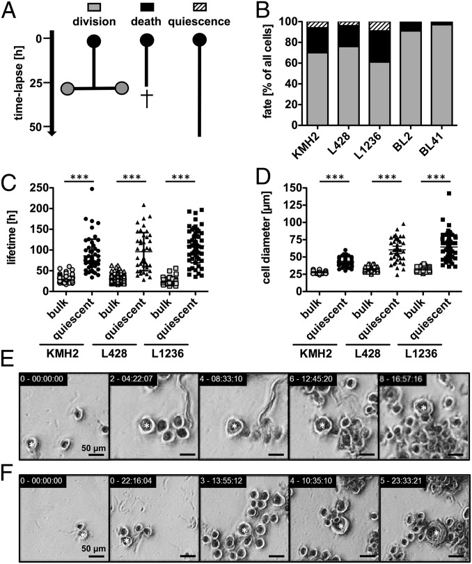
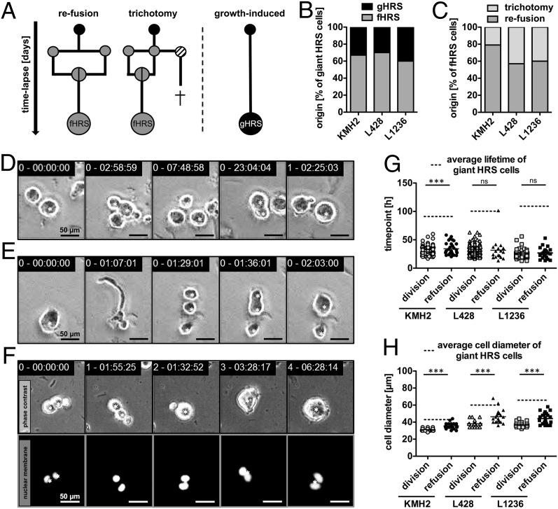
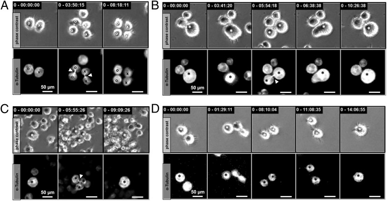
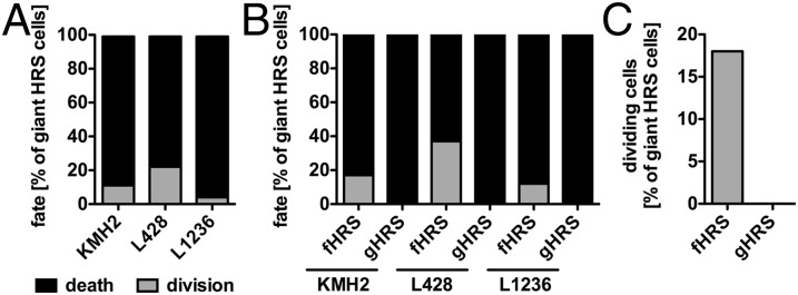
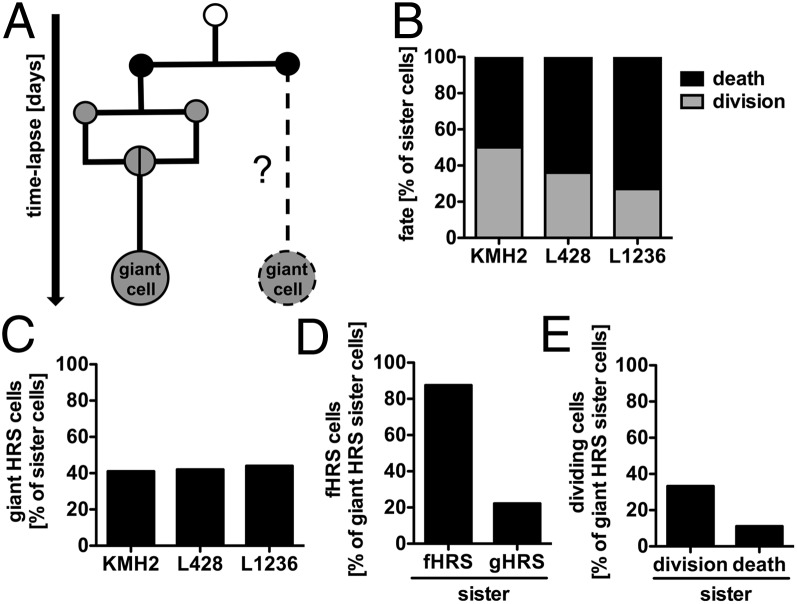
Multinucleated Reed-Sternberg (RS) cells are pathognomonic for classical Hodgkin lymphoma (HL), and their presence is essential for diagnosis. How these giant tumor cells develop is controversial, however. It has been postulated that RS cells arise from mononucleated Hodgkin cells via endomitosis. Conversely, continuous single-cell tracking of HL cell lines by long-term time-lapse microscopy has identified cell fusion as the main route of RS cell formation. In contrast to growth-induced formation of giant Hodgkin cells, fusion of small mononuclear cells followed by a size increase gives rise to giant RS cells. Of note, fusion of cells originating from the same ancestor, termed re-fusion, is seen nearly exclusively. In the majority of cases, re-fusion of daughter cells is preceded by incomplete cytokinesis, as demonstrated by microtubule bonds among the cells. We confirm at the level of individual tracked cells that giant Hodgkin and RS cells have little proliferative capacity, further supporting small mononuclear Hodgkin cells as the proliferative compartment of the HL tumor clone. In addition, sister cells show a shared propensity for re-fusion, providing evidence of early RS cell fate commitment. Thus, RS cell generation is related neither to cell fusion of unrelated Hodgkin cells nor to endomitosis, but rather is mediated by re-fusion of daughter cells that underwent mitosis. This surprising finding supports the existence of a unique mechanism for the generation of multinuclear RS cells that may have implications beyond HL, given that RS-like cells are frequently observed in several other lymphoproliferative diseases as well.


Conflict of interest statement

The authors declare no conflict of interest.

Figures

Fig. 1.
Fig. 1.
HL cell lines contain rare giant and long-lived HRS cells. Individual cells of HL cell lines KMH2, L428, and L1236 and their progeny were followed with time-lapse microscopy-based cell tracking for 10–12 d, and pedigrees were generated. (A and B) The cell fate of all seeded cells (n = 107–146; generation 0) within the first 50 h was analyzed. (A) Schematic diagram of the identified fates in HL cell lines. Quiescence is defined as cell survival of longer than 50 h without division. (B) Cell fate distribution in HL and BL cell lines (BL2 and BL41), which served as negative controls without a quiescent population. Results for subsequent generations 1 and 2 are shown in Fig. S1A. (C and D) Quiescent cells (n = 36–54, all generations) demonstrated a longer lifetime (C) and larger cell diameter (D) compared with the corresponding bulk population. (E and F) Example images showing representative HRS cell development in the L428 cell line. Shown are the lifespan of a long-lived, quiescent HRS cell (E; 8.5 d; Movie S1) and growth of a normal-sized cell becoming a giant HRS cell (F; 2.2-fold size increase). Stars in E and F designate cells tracked over time. Equivalent examples of KMH2 and L1236 cells are shown in Fig. S1 C and D.
Fig. 2.
Fig. 2.
RS cells arise primarily from re-fusion of daughter cells. Development of giant HRS cells was corroborated by time-lapse microscopy-based cell tracking of HL cell lines KMH2, L428, and L1236. (A–C) Continuous HRS cell tracking (n = 36–54) identified re-fusion of daughter cells as a prominent route of giant cell formation. Occasionally, cell fusion was present as a so-called trichotomy, meaning a division into three cells with subsequent fusion of two of the three daughter cells. Most of the remaining nonfused daughter cells died. (A) Schematic diagram of observed routes of giant HRS cell formation. (B) Frequency of fHRS and gHRS cells. (C) Frequency of fHRS cells originating via trichotomy. (D and E) Examples of L1236 cell division and subsequent re-fusion (D; Movie S2) and trichotomy (E) demonstrating the division into three cells with fusion of two cells leading to two daughter cells of unequal size (Movie S3). Equivalent examples from HL cell lines KMH2 and L428 are shown in Fig. S2 A and B. (F) Tracking of the development of a KMH2 fHRS cell with nuclear fluorescence, focusing on cell fusion (Movie S5). An example of a gHRS cell is shown in Fig. S3A. (G) Comparison of the generation time of dividing cells with the division time of giant HRS cells before a cell fusion event. (H) Average cell size of the bulk population plotted against the cell diameter of giant HRS cells before cell fusion occurred. The tracked cells are denoted by a star in DF.
Fig. 3.
Fig. 3.
Incomplete cytokinesis precedes re-fusion of HRS daughter cells. (A) Proliferating HRS cells with fluorescent microtubules (RFP-tubulin) demonstrated midbody assembly during cytokinesis. Mitosis is completed by disassembly of the microtubule connection between the daughter cells. (B) In 83% of the 36 observed fHRS cell formations, daughter cells remained connected via the midbody until re-fusion occurred (Movie S7). (C) Multidaughter divisions displayed a three-part midbody connection. (D) In some cases (∼17%), cytokinesis was complete before cells re-fused, as demonstrated by disassembly of the connecting midbody (Movie S8). Tracked cells are denoted by a circle or cross in A and by a star in B, C, and D. The midbody is indicated by an arrowhead.
Fig. 4.
Fig. 4.
Re-fusion events predict the residual proliferation potential of RS cells. The fate of individual giant HRS cells (n = 36–54) was determined by continuous tracking of HL cell lines KMH2, L428, and L1236. (A) Only cells with a distinct cell fate until the end of the observation period (death or division) were subjected to analysis. (B) Giant HRS cells were grouped by dependency of their origin, and cell fate was reassessed. (C) Highlighted proliferation potentials of fHRS and gHRS cells.
Fig. 5.
Fig. 5.
RS cell development is committed in ancestor generations. (A) Pedigree analyses revealed the cell behavior and fate of sister cells of giant HRS cells (n = 13–28) for the KMH2, L428, and L1236 cell lines. (B and C) Continuous single-cell tracking of HRS sister cells determined cell fate (B) and the incidence of giant cell development (C). (D) Likelihood of detecting fHRS cell development in fHRS or gHRS sister cells. (E) Likelihood of identifying giant HRS cell division when the corresponding sister cell is a dividing or dying giant cell.

References

    1. Hodgkin T. On some morbid appearances of the absorbent glands and spleen. Med Chir Trans. 1832;17:68–114. - PMC - PubMed
    1. Drexler HG. Recent results on the biology of Hodgkin and Reed-Sternberg cells, I: Biopsy material. Leuk Lymphoma. 1992;8(4-5):283–313. - PubMed
    1. Sternberg C. Über eine eigenartige unter dem bilde der pseudoleukämie verlaufende tuberkulose des lymphatischen apparates. Zeitschrift für Heilkunde. 1898;19:21–90.
    1. Reed DM. On the pathological changes in Hodgkin’s disease, with special reference to its relation to tuberculosis. Johns Hopkins Hosp Rep. 1902;10:133–196.
    1. Küppers R, Schwering I, Bräuninger A, Rajewsky K, Hansmann ML. Biology of Hodgkin’s lymphoma. Ann Oncol. 2002;13(Suppl 1):11–18. - PubMed

Publication types