. 2013 Nov 26;8(11):e80805.
doi: 10.1371/journal.pone.0080805. eCollection 2013.

Dyskerin localizes to the mitotic apparatus and is required for orderly mitosis in human cells

Affiliations

Dyskerin localizes to the mitotic apparatus and is required for orderly mitosis in human cells

Faizan Alawi et al. PLoS One. .

Abstract

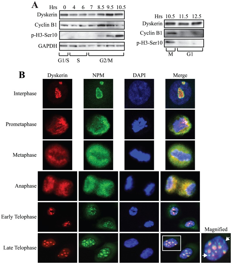
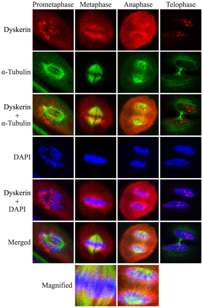
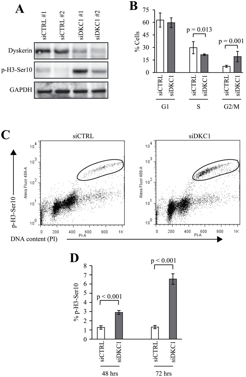
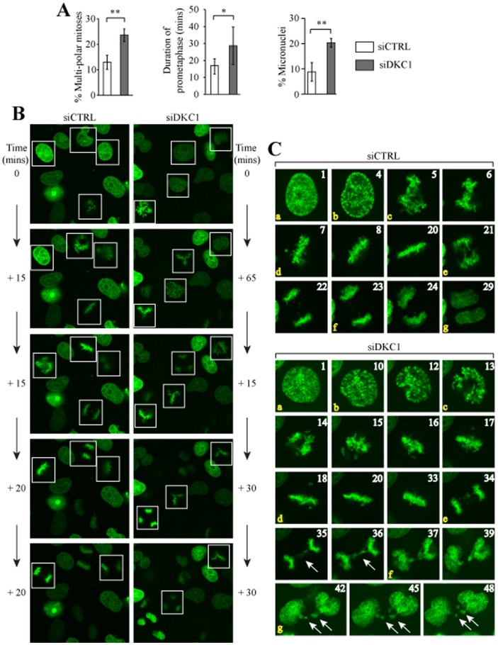
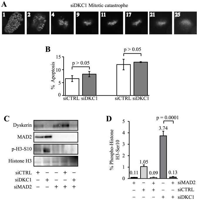
Dyskerin is a highly conserved, nucleolar RNA-binding protein with established roles in small nuclear ribonucleoprotein biogenesis, telomerase and telomere maintenance and precursor rRNA processing. Telomerase is functional during S phase and the bulk of rRNA maturation occurs during G1 and S phases; both processes are inactivated during mitosis. Yet, we show that during the course of cell cycle progression, human dyskerin expression peaks during G2/M in parallel with the upregulation of pro-mitotic factors. Dyskerin redistributed from the nucleolus in interphase cells to the perichromosomal region during prometaphase, metaphase and anaphase. With continued anaphase progression, dyskerin also localized to the cytoplasm within the mid-pole region. Loss of dyskerin function via siRNA-mediated depletion promoted G2/M accumulation and this was accompanied by an increased mitotic index and activation of the spindle assembly checkpoint. Live cell imaging further revealed an array of mitotic defects including delayed prometaphase progression, a significantly increased incidence of multi-polar spindles, and anaphase bridges culminating in micronucleus formation. Together, these findings suggest that dyskerin is a highly dynamic protein throughout the cell cycle and increases the repertoire of fundamental cellular processes that are disrupted by absence of its normal function.


Conflict of interest statement

Competing Interests: The authors have declared that no competing interests exist.

Figures

Figure 1
Figure 1. Dyskerin expression peaks during G2/M and localizes to distinct compartments throughout mitosis.
A, UM-SCC1 cells were synchronized at the beginning of S phase using a double thymidine block. Western blot showing expression of dyskerin at various times after release from the block. Cyclin B1 and p-H3-Ser10 expression were used to highlight G2/M. GAPDH was used as a loading control. B, Dyskerin showed strong co-localization with NPM throughout prometaphase, metaphase and anaphase. Dyskerin partially co-localized with NPM during interphase and telophase. During telophase, nucleoplasmic NPM foci devoid of dyskerin are also seen. The boxed nucleus is magnified to show detail. NPM foci are indicated by arrows. One representative OKF6-TERT2 cell is shown at each mitotic stage.
Figure 2
Figure 2. Dyskerin is mostly excluded from the mitotic spindle.
Co-localization of dyskerin with α-tubulin was either weak or inconsistently observed. Thus, specific localization of dyskerin with the mitotic spindle could not be reliably ascertained by indirect immunofluorescence. The merged metaphase and anaphase panels are magnified to show detail. One representative OKF6-TERT2 cell is shown at each stage. Dyskerin (red), α-tubulin (green), DAPI (blue).
Figure 3
Figure 3. Dyskerin depletion leads to an increased mitotic index.
A, After 72 hrs, dyskerin levels were decreased in HeLa cells by more than 80% following transfection with two distinct siRNAs directed against DKC1 (siDKC1) relative to cells transfected with non-target siRNAs (siCTRL). This was accompanied by activation of p-H3-Ser 10 expression. siDKC1 #1 – custom-designed siRNA pool (as described in Materials and Methods), siDKC1 #2 – pre-designed siRNA pool (Dharmacon). B, Loss of dyskerin led to a small but statistically significant increase in G2/M accumulation as measured by flow cytometry. C, The G2/M accumulation was partially attributable to an increase in the mitotic fraction of dyskerin-depleted cells 72 hrs after siRNA transfection as measured by anti-p-H3-ser 10-Alexa Fluor® 488 and propidium iodide labeling (circled populations). D, The mitotic fractions were quantitated 48 and 72 hrs after siRNA transfection. Experiments in panels B–D were performed using siDKC1 #1.
Figure 4
Figure 4. Loss of dyskerin delays prometaphase, disrupts spindle formation and promotes micronucleus formation.
HeLa-H2B-GFP cells were transfected with siDKC1 #1 (siDKC1) or control siRNA (siCTRL). After 48 hrs, images were captured in real-time every five minutes for 16 hrs. A, Quantitation of multi-polar mitoses as a percentage of the total number of mitoses counted; siCTRL (N = 110), siDKC1 (N = 148). Quantification of prometaphase duration and micronuclei formation; N = 30 for each condition. *p<0.0001, **p<0.002. B, Random fields demonstrating four siCTRL and siDKC1 cells undergoing mitosis (white boxes), respectively, are shown for illustrative purposes. The panels at the top are arbitrarily assigned as Time 0. Each panel going down represents the same field and shows the cells as they progressed from prophase through to anaphase. The times listed adjacent to each panel represent the amount of time it took to reach the stage shown, as measured by the number of consecutive image captures multiplied by 5 mins per image capture. All four mitoses in the siDKC1 field yielded tri-polar mitoses. C, Single representative cells are shown as they progressed from prophase to cytokinesis. a: The panels assigned as ‘1’ represent the cells during prophase. The numbers in the other panels represent the image capture number subsequent to image ‘1’. b: siCTRL panel ‘4’ and siDKC1 panel ‘10’ represent late prophase during which the chromatin can be seen condensing. c: prometaphase begins; d: metaphase begins; e: anaphase; f: telophase; g: cytokinesis. The duration of time needed for this siCTRL cell to progress from b (late prophase) – f (telophase) was 95 mins. The corresponding siDKC1 cell required 130 mins. Lagging chromosomes and a chromatin bridge are seen during anaphase (panels ‘35’ and ‘36’, arrows) in the siDKC1 cell. Micronuclei are also evident following cytokinesis (panels ‘42’-‘48’, arrows).
Figure 5
Figure 5. Loss of dyskerin triggers the spindle assembly checkpoint.
A, Representative Hela-H2B-GFP siDKC1 cell progressing from prophase to mitotic catastrophe. The numbers shown in each panel represent the image capture number subsequent to image ‘1’. B, Three and six days after HeLa transfection apoptosis was assessed using Annexin V staining and flow cytometry. Error bars denote the standard deviations derived from three independent transfections at each time point. One representative experiment is shown. C, HeLa cells were transfected with CTRL, DKC1 and/or MAD2 siRNAs and harvested 72 hrs later. Immunoblot analysis showed almost complete absence of MAD2 and p-H3-Ser 10 expression in MAD2- depleted cells. D, In parallel, the cells were fixed and the mitotic fractions were assessed by flow cytometry.

References

    1. Meier UT (2006) How a single protein complex accommodates many different H/ACA RNAs. Trends Biochem Sci 31: 311–315. - PMC - PubMed
    1. Ruggero D, Grisendi S, Piazza F, Rego E, Mari F, et al. (2003) Dyskeratosis congenita and cancer in mice deficient in ribosomal RNA modification. Science 299: 259–262. - PubMed
    1. Lafontaine DL, Bousquet-Antonelli C, Henry Y, Caizergues-Ferrer M, Tollervey D (1998) The box H + ACA snoRNAs carry Cbf5p, the putative rRNA pseudouridine synthase. Genes Dev 12: 527–537. - PMC - PubMed
    1. Giordano E, Peluso I, Senger S, Furia M (1999) minifly, a Drosophila gene required for ribosome biogenesis. J Cell Biol 144: 1123–1133. - PMC - PubMed
    1. Mitchell JR, Wood E, Collins K (1999) A telomerase component is defective in the human disease dyskeratosis congenita. Nature 402: 551–555. - PubMed

Publication types

LinkOut - more resources