. 2013 Nov 26;8(11):e81306.
doi: 10.1371/journal.pone.0081306. eCollection 2013.

Identification of a novel zinc metalloprotease through a global analysis of Clostridium difficile extracellular proteins

Affiliations

Identification of a novel zinc metalloprotease through a global analysis of Clostridium difficile extracellular proteins

Valeria Cafardi et al. PLoS One. .

Abstract

Clostridium difficile is a major cause of infectious diarrhea worldwide. Although the cell surface proteins are recognized to be important in clostridial pathogenesis, biological functions of only a few are known. Also, apart from the toxins, proteins exported by C. difficile into the extracellular milieu have been poorly studied. In order to identify novel extracellular factors of C. difficile, we analyzed bacterial culture supernatants prepared from clinical isolates, 630 and R20291, using liquid chromatography-tandem mass spectrometry. The majority of the proteins identified were non-canonical extracellular proteins. These could be largely classified into proteins associated to the cell wall (including CWPs and extracellular hydrolases), transporters and flagellar proteins. Seven unknown hypothetical proteins were also identified. One of these proteins, CD630_28300, shared sequence similarity with the anthrax lethal factor, a known zinc metallopeptidase. We demonstrated that CD630_28300 (named Zmp1) binds zinc and is able to cleave fibronectin and fibrinogen in vitro in a zinc-dependent manner. Using site-directed mutagenesis, we identified residues important in zinc binding and enzymatic activity. Furthermore, we demonstrated that Zmp1 destabilizes the fibronectin network produced by human fibroblasts. Thus, by analyzing the exoproteome of C. difficile, we identified a novel extracellular metalloprotease that may be important in key steps of clostridial pathogenesis.


Conflict of interest statement

Competing Interests: Valeria Cafardi, Massimiliano Biagini, Manuele Martinelli, Rosanna Leuzzi, Nathalie Norais, Maria Scarselli, Davide Serruto and Meera Unnikrishnan are employed by Novartis Vaccines and Diagnostics, which partly funded this research. This does not alter the authors' adherence to all the PLOS ONE policies on sharing data and materials.

Figures

Figure 1
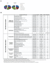
Figure 1. Identification of extracellular proteins from Clostridium difficile culture supernatants.
(A) Family distribution of putative surface/released proteins detected in 630 and R20291 culture supernatants. Proteins identified have been divided in families based primarily on the presence of motifs for association to the peptidoglycan and secondly on the presence of conserved functional domains. (B) Proteins detected by LC-MS/MS in C. difficile 630 and R20291 culture supernatants. Presence of a signal peptide motif was predicted with SignalP 4.0 using the 630 amino acid sequence (except for CDR20291_2278, for which a sequence with amino acid identity >40% is not present in 630). Cellular localization was predicted by PSORTb 3.0 using the 630 amino acid sequence (except for CDR20291_2278). Detection in 630 or R20291 culture supernatants is indicated by ‘yes/no’.
Figure 2
Figure 2. Cellular localization of C. difficile 630 proteins detected by LC-MS/MS.
Immunoblotting analysis of cell and supernatant fractions prepared from exponential phase cultures with antisera generated against CD630_01830, CD630_23650, CD630_02370 (FliD) and CD630_28300. Cell fractions: total extract (TE), protoplast (P), mutanolysin extract (ME), S-layer extract (SL). Supernatant fractions: total supernatant (TS), supernatant after ultracentrifugation (US), ultracentrifugation pellet (UP). 10 or 50 ng of each recombinant protein (RP) was loaded as control.
Figure 3
Figure 3. Zmp1 is a zinc-dependent protease with fibrinogen- and fibronectin-cleaving activity.
(A) Zmp1 shares sequence similarity with the C-terminal ATLF catalytic domain of anthrax lethal factor. Residues previously involved in the zinc proteolytic activity (indicated in bold) are conserved. (B) Time-dependent proteolytic activity of recombinant Zmp1 on fibronectin. 1µM fibronectin from human plasma was incubated for 24 h at 37°C with 7.7 µM Zmp1 in the presence of 0.5 mM ZnCl2. At 0, 1, 3, 6 and 24 h after incubation, 1 µg of fibronectin was analyzed by SDS-PAGE followed by Coomassie-blue staining. Integrity of fibronectin in the absence of Zmp1 was verified after 24 h of incubation in the same conditions. (C) Concentration-dependent activity of Zmp1 on fibronectin was observed after incubation of different concentrations of Zmp1 with 1 µM of fibronectin for 24 h at 37°C in the presence of 0.5 mM ZnCl2. 1 µg of fibronectin was analyzed by SDS-PAGE and Coomassie-blue staining. (D) Time-dependent proteolytic activity of recombinant Zmp1 on fibrinogen. 1 µM fibrinogen was incubated for 24 h at 37°C with 1 µM Zmp1 in the presence of 0.5 mM ZnCl2. At 0, 1, 3, 6 and 24 h after incubation, 5 µg of fibrinogen was analyzed by SDS-PAGE followed by Coomassie-blue staining. Integrity of fibrinogen in the absence of Zmp1 was verified after 24 h of incubation in the same conditions. (E) Concentration-dependent activity of Zmp1 on fibrinogen was observed after incubation of different concentrations of Zmp1 with 1 µM of substrate for 6 h at 37°C in the presence of 0.5 mM ZnCl2. 5 µg of fibrinogen was analyzed by SDS-PAGE and Coomassie-blue staining.
Figure 4
Figure 4. Impaired fibronectin and fibrinogen-cleaving activities of the E143A and H146A Zmp1 mutants.
(A) Differential scanning fluorimetry of wild type, E143A and H146A recombinant proteins in the absence or presence of 1:16 Zn2+. The minimum of the derivative is the melting temperature. Shifts in melting curves for the wild type and mutant proteins are indicated with arrows. (B) NMR analysis of wild type, E143A and H146A. 1H15N HSQC spectrum of apo (black) and in the presence of zinc (red) of Zmp1 wild type, H146A and E143A. (C) Decreased ability of E143A and H146A Zmp1 mutants to cleave fibronectin. 1 µM fibronectin from human plasma was incubated for 24 h at 37°C with no Zmp1 (control) or with 7.7 µM of wild type (WT), E143A or H146A Zmp1 in the presence of 0.5 mM ZnCl2. 1 µg of fibronectin was analyzed by SDS-PAGE and Coomassie-blue staining. (D) Inability of E143A and H146A Zmp1 mutants to cleave fibrinogen. 1 µM fibrinogen was incubated for 24 h at 37°C with 1 µM of wild type (WT), E143A or H146A Zmp1 in the presence of 0.5 mM ZnCl2. 1 µg of fibrinogen was analyzed by SDS-PAGE and Coomassie-blue staining.
Figure 5
Figure 5. Proteolytic activity of Zmp1 protein on native fibronectin produced by cultured human fibroblasts.
(A) IMR-90 human fibroblasts were incubated with 11.6 µM of wild type, E143A or H146A Zmp1 protein for 16 h as described in Methods. Fibronectin was labeled using anti-fibronectin followed by Alexa568-conjugated secondary antibodies (red) and nuclei were stained with DAPI (blue). Control cells were incubated with an equivalent volume of buffer for the same time period. Destabilization of fibronectin was observed upon treatment of cells with Zmp1, as also highlighted in the inset. The images shown are representative of 3 independent experiments. (B) Immunoblotting analysis of culture supernatants from IMR-90 cells treated with 11.6 µM of wild type, E143A or H146A Zmp1 protein for 16 h. 100 µl undiluted supernatants from each well were probed with anti fibronectin.

References

    1. Rupnik M, Wilcox MH, Gerding DN (2009) Clostridium difficile infection: new developments in epidemiology and pathogenesis. Nat Rev Microbiol 7: 526-536. doi:10.1038/nrmicro2164. PubMed: 19528959. - DOI - PubMed
    1. Kuipers EJ, Surawicz CM (2008) Clostridium difficile infection. Lancet 371: 1486-1488. doi:10.1016/S0140-6736(08)60635-2. PubMed: 18456087. - DOI - PubMed
    1. Warny M, Pepin J, Fang A, Killgore G, Thompson A et al. (2005) Toxin production by an emerging strain of Clostridium difficile associated with outbreaks of severe disease in North America and Europe. Lancet 366: 1079-1084. doi:10.1016/S0140-6736(05)67420-X. PubMed: 16182895. - DOI - PubMed
    1. O'Connor JR, Johnson S, Gerding DN (2009) Clostridium difficile infection caused by the epidemic BI/NAP1/027 strain. Gastroenterology 136: 1913-1924. doi:10.1053/j.gastro.2009.02.073. PubMed: 19457419. - DOI - PubMed
    1. Surawicz CM, Alexander J (2011) Treatment of refractory and recurrent Clostridium difficile infection. Nat Rev Gastroenterol Hepatol 8: 330-339. doi:10.1038/nrgastro.2011.59. PubMed: 21502971. - DOI - PubMed

Publication types

MeSH terms

LinkOut - more resources