. 2013 Nov 26;8(11):e81695.
doi: 10.1371/journal.pone.0081695. eCollection 2013.

Respiratory syncytial virus induced type I IFN production by pDC is regulated by RSV-infected airway epithelial cells, RSV-exposed monocytes and virus specific antibodies

Affiliations

Respiratory syncytial virus induced type I IFN production by pDC is regulated by RSV-infected airway epithelial cells, RSV-exposed monocytes and virus specific antibodies

Marcel A Schijf et al. PLoS One. .

Abstract

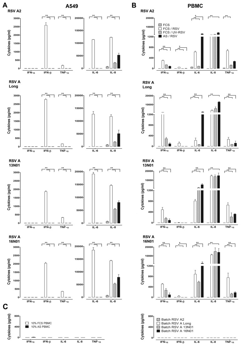
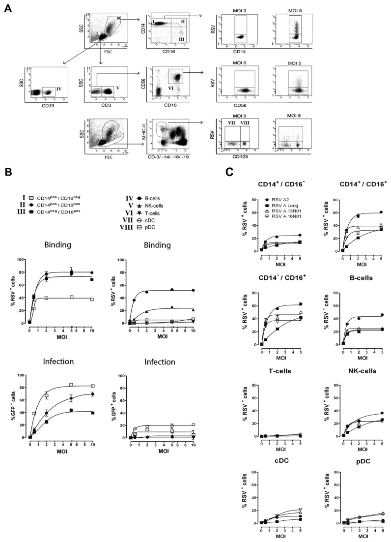
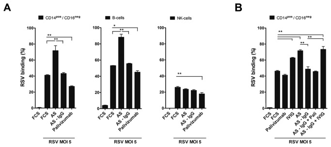
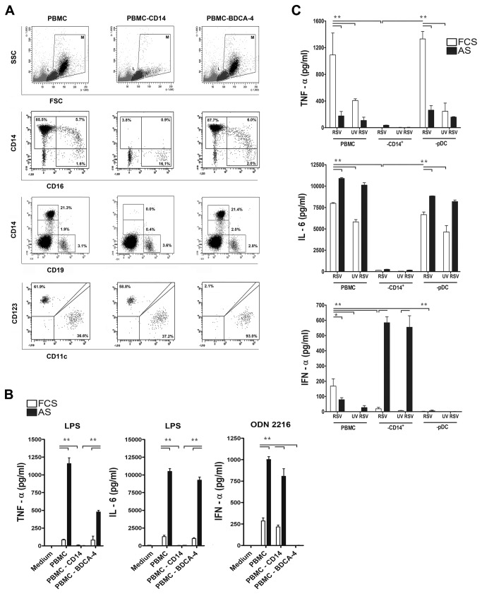
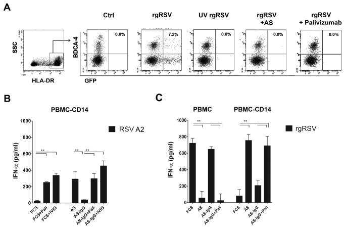
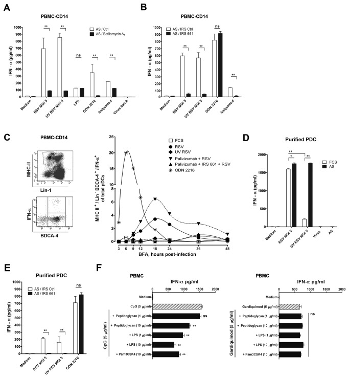
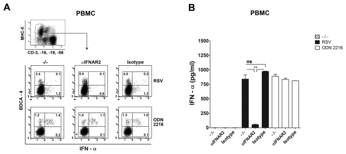
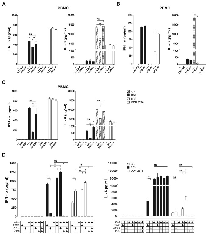
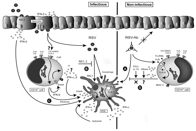
Innate immune responses elicited upon virus exposure are crucial for the effective eradication of viruses, the onset of adaptive immune responses and for establishing proper immune memory. Respiratory syncytial virus (RSV) is responsible for a high disease burden in neonates and immune compromised individuals, causing severe lower respiratory tract infections. During primary infections exuberant innate immune responses may contribute to disease severity. Furthermore, immune memory is often insufficient to protect during RSV re-exposure, which results in frequent symptomatic reinfections. Therefore, identifying the cell types and pattern recognition receptors (PRRs) involved in RSV-specific innate immune responses is necessary to understand incomplete immunity against RSV. We investigated the innate cellular response triggered upon infection of epithelial cells and peripheral blood mononuclear cells. We show that CD14(+) myeloid cells and epithelial cells are the major source of IL-8 and inflammatory cytokines, IL-6 and TNF-α, when exposed to live RSV Three routes of RSV-induced IFN-α production can be distinguished that depend on the cross-talk of different cell types and the presence or absence of virus specific antibodies, whereby pDC are the ultimate source of IFN-α. RSV-specific antibodies facilitate direct TLR7 access into endosomal compartments, while in the absence of antibodies, infection of monocytes or epithelial cells is necessary to provide an early source of type I interferons, required to engage the IFN-α,β receptor (IFNAR)-mediated pathway of IFN-α production by pDC. However, at high pDC density infection with RSV causes IFN-α production without the need for a second party cell. Our study shows that cellular context and immune status are factors affecting innate immune responses to RSV. These issues should therefore be addressed during the process of vaccine development and other interventions for RSV disease.


Conflict of interest statement

Competing Interests: The authors have declared that no competing interests exist.

Figures

Figure 1
Figure 1. Cell specific innate immune response to RSV.
Cytokine responses upon RSV exposure by lung epithelial cell line A549 (A) and human PBMC (B). Cells were co-cultured with the virus strains: RSV A2, RSV Long strain or 2 recent subtype A isolates, 13NO1 and 16NO1 at MOI 5. In addition to live RSV, the response to UV-inactivated and neutralized RSV in autologous serum (AS) was measured. After 20 hrs. incubation, cytokines in supernatant were measured by multiplex immunoassay and ELISA. In figure 1A the data represent mean values from duplicate experiments using human serum from four different donors. Figure 1B shows the cytokine responses in human PBMC exposed to RSV A2, representing the mean values measured in 6 different donors. For the other virus strains, means for four different donors are depicted whereby similar results were obtained in duplicate experiments. (C) Control measurements of cytokines produced in PBMC cultured in the presence of medium containing 10% FCS or autologous serum (AS, left) and cytokine measurements in the virus batches used in panels A and B (right). Data represent the mean ± SEM and were analyzed using two way ANOVA followed by a Bonferroni post-test, *P< 0.05, **P <0.01.
Figure 2
Figure 2. Binding efficiency of RSV is cell type specific and does not correlate with the susceptibility of the cells to infection.
Binding and infection of rgRSV224 to different cell types in human PBMC was investigated. (A) FACS gating strategy for CD14+/CD16neg., CD14+/CD16+ and CD14neg./CD16+ monocytes (in M: monocyte gate), CD19+ B-cells, CD3+ T-cells and CD3neg.CD16+CD56+ NK cells (in L: Lymphocyte gate). Dendritic cell populations were identified by CD3, CD14, CD16, CD19neg. (Linneg.), MHC-IIhigh, and either CD123 (cDC) or CD123+ (pDC) (in the total live cell gate, M+L). (B) Binding of RSV to specific cell types was measured after 1 hour incubation at 4°C with polyclonal antibodies against RSV (upper figures). RSV infection was determined after 24 hrs. incubation at 37°C by the percentage of GFP expressing cells within a specific cell population (lower figures). (C) Cell type specific binding characteristics of 4 RSV A strains. Binding of RSV A2, RSV A Long, and clinical RSV A isolates 13N01 and 16N01 to specific cell types in PBMC was visualized after 1 hour incubation at 4°C. Binding was detected using polyclonal antibodies against RSV. Data represent the percentage of RSV positive cells within a population and are expressed as mean ± SEM of triplicate measurements within 1 donor. Experiments were performed in 5 different donors with similar results.
Figure 3
Figure 3. Virus specific polyclonal antibodies in human serum increase RSV binding to monocytes and B cells.
(A) The effect of autologous serum, IgG-depleted serum and palivizumab in FCS on binding of RSV to monocytes, B cells and NK cells was measured after 1 hour incubation at 4°C. (B) Polyclonal antibodies (IVIG) increase binding of RSV to CD14+/CD16neg. cells. Data shown represent the mean ± SEM of triplicate measurements in two different donors (for A and B) and were analyzed using the Kruskal-Wallis test followed by Dunn's Multiple Comparison analysis, *P< 0.05, **P <0.01. Experiments were performed in 3 additional donors with similar results.
Figure 4
Figure 4. CD14+ cells are needed for the inflammatory cytokine response against RSV in PBMC cultures.
(A) Confirmation of specific depletion of CD14+ and BDCA-4+ cells by FACS. M: monocyte gate, L: lymphocyte gate. Depletion of CD14+ cells leaves B cells (in M+L, CD19+ third row) and CD16+ monocyte/DC (in M, CD14neg., CD16+, 2nd row), cDC (in M+L, lineageneg., MHCII+, CD11c+, 4rd row) and pDC (in M+L, lineageneg., MHCII+, BDCA-4+, 4rd row) compartments intact. Depletion of BDCA-4+ cells removes pDC but does not affect CD14+CD16neg., CD14+CD16+, CD14neg.CD16+ cells in the M gate (2nd row), nor B cells (3rd row) and cDC (4rd row). The contribution of single cell types to anti-RSV cytokine responses in PBMC cultures was evaluated by depletion of specific cell populations. Cytokines in supernatant from the remaining cell populations were measured after 20 hrs. exposure to RSV or UV-RSV in the presence or absence of autologous serum. (B) Depletion of CD14+ cells and BDCA-4+ cells was confirmed by stimulation of depleted cell populations by TLR ligands, ultra-pure LPS to confirm the absence of TLR4+ (monocytes) and ODN 2216 to confirm the absence of TLR9+ (pDC). (C) PBMC, CD14 monocyte-depleted PBMC, or pDC-depleted PBMC were cultured with live and UV inactivated RSV (A2, MOI 5) in the presence or absence of 10% autologous serum. Data represent the mean ± SEM of 3 measurements in 1 donor and were analyzed using one way ANOVA followed by a Bonferroni post-test, *P< 0.05, **P <0.01. Experiments were performed in 3 different donors with similar results.
Figure 5
Figure 5. RSV-specific antibodies inhibit RSV-induced IFN-α production in PBMC, but enhance IFN-α production in CD14+ cell depleted PBMC.
(A) Inhibition of RSV infection in pDC. Lineageneg., MHC-IIhigh, BDCA-4+ pDC are partially infected with rgRSV224 after a period of 20 hours. Infection is blocked after UV inactivation, after neutralization in 10% fresh human serum or in 5µg/ml Palivizumab. Similar results were obtained with sera from all donors used during our studies. IFN-α in supernatant of RSV-A2 exposed PBMC-CD14+ cell cultures (B) and rgRSV224 exposed PBMC-CD14+ cells or PBMC, (C) was measured after 20 hrs. The role of virus specific antibodies on the cytokine response was tested by removing IgGs from AS with protein G Sepharose® beads and reconstitution with 2 mg/ml IVIG (B) or 5 µg/ml palivizumab (B, C). Experiments represent the mean ± SEM of experiments performed in 4 different donors and were analyzed using one way ANOVA followed by a Bonferroni post-test, **P <0.01.
Figure 6
Figure 6. CD14+ monocytes inhibit IFN-α production triggered by Ab-RSV via TLR7 in pDC.
(A) IFN-α production in CD14+ cell depleted PBMCs induced by Ab-RSV complexes, TLR9 ligand ODN 2216 and TLR7 ligand imiquimod is decreased by blocking endosomal acidification with 50nM Bafilomycin A1. (B) Ab-RSV-induced IFN-α production in CD14+ cell depleted PBMCs was abrogated in the presence of immune regulatory sequence (IRS) 661 (1.4 µM) a specific blocking agent for endosomal TLR7 and not by a scrambled control nucleotide. (C) pDC are the source of Ab-RSV induced, TLR7 mediated production of IFN-α, as shown by intracellular staining for IFN-α in Lineage (CD3neg., CD14neg., CD19neg., CD16neg., CD56neg., Lin-1), MHC-IIhigh, BDCA-4+ cells. The inhibitor brefeldin A was added at different time points post infection (the time points when BFA was added are given on the X-axis). Cytokines were allowed to accumulate for 10 hrs. after addition of BFA. (D) Purified pDC (obtained by negative selection removing CD3+, CD19+ and CD16+ cells from fresh PBMC, followed by FACS purification of the BDCA-4+ cell population, which resulted in > 95% pure pDC) produce IFN-α upon infection with RSV. This response is abrogated after UV inactivation of RSV. In AS, both live RSV and UV-inactivated RSV induced IFN-α production to a similar extent. One representative experiment out of two performed with pDC isolated from two different donors is shown. (E) IFN-α production by Ab-RSV in purified pDC is blocked by IRS661 (1.4µM). (F) TLR1,-2 (PAM3CSK4, Peptidoglycan) and TLR4 (LPS) ligands suppress TLR9-triggered (ODN 2216) IFN-α production, but do not affect TLR7 (Gardiquimod) induced IFN-α production. All data represent mean ± SEM of triplicate measurements within 1 donor and analyzed using one way ANOVA followed by a Bonferroni post-test. ns not significant, *P< 0.05, **P <0.01. Experiments were performed in 3 different donors with similar results.
Figure 7
Figure 7. IFN-α production induced by live RSV in PBMC depends on IFNAR signalling.
PBMC were exposed to live RSV in the presence of interferon-α/β receptor (IFNAR) blocking antibody (5µg/ml), isotype control Ab or no Ab. Levels of IFN-α were determined via intracellular staining (A) or in 20hrs. supernatant by ELISA (B). IFN-α was trapped intracellular by BFA treatment initiated 6 hrs. after RSV infection, or at t=0 for the ODN 2216 control stimulus, because of different kinetics of anti-viral and ODN elicited IFN-α response. For both stimuli, intracellular staining for IFN-α was performed in CD3neg., CD16neg., CD19neg., CD56neg., MHCII+, BDCA-4+ cells after 10 hrs. BFA treatment. Experiments were performed in 3 different donors with similar results. Data represent the mean ± SEM of triplicate measurements within 1 donor and were analyzed using one way ANOVA followed by a Bonferroni post-test. ns not significant, **P <0.01.
Figure 8
Figure 8. IFN-α production induced by live RSV in pDC depends on CD14.
The role of the TLR4/CD14/MD2 complex in the IFN-α and IL-6 response by PBMC after RSV infection (strain A2, MOI 5) was investigated (A) with a blocking monoclonal antibody specific for human TLR4 (20µg/ml), (B) the MD2 antagonist lipopolysaccharide from the bacterium Rhodobacter sphaeroides (1µg/ml) or (C) via neutralization of CD14 with monoclonal antibody MY4 (10µg/ml). (A-C) Cytokine responses were measured by ELISA. Control stimuli; LPS: 10ng/ml, ODN: 5µg/ml. (D) A549 epithelial cells were co-cultured with PBMC, CD14+ cell depleted PBMC, pDC depleted PBMC, or CD14+ and BDCA-4+ double depleted PBMC, in the presence of RSV at MOI 5, ODN 2216 or no stimulus, for a period of 20 hrs. IL-6 and IFN-α production were measured by ELISA. Experiments were performed in 3 different donors with similar results. Data represent the mean ± SEM of triplicate measurements within 1 donor and were analyzed using one way ANOVA followed by a Bonferroni post-test. ns not significant, *P<0.05, **P<0.01.
Figure 9
Figure 9. Cellular cross talk and the role of antibodies during IFN-α production.
pDC produce IFN-α via multiple pathways. i: TLR7 activation after Ab-mediated uptake of RSV (A), ii: via infection with RSV in purified pDC (B) or iii: indirect via IFNAR-mediated signalling triggered by IFN-β produced by RSV-infected A549 (as a model for airway epithelium) or RSV-exposed CD14+ cells (C). In the context of PBMC the presence of CD14+ monocytes suppresses the production of IFN-α by pDC upon exposure to Ab-RSV complexes. (D). The mechanism of the suppression by monocytes of this TLR7-mediated response elicited by Ab-RSV complexes in pDC remains unresolved, but appears not to be mediated via PG’s and IL-10, mediators that suppress TLR9 induced IFN-α production when multiple PAMPS in bacterial lysates simultaneously trigger TLR9 and TLR2/4 responses in PBMC.

Similar articles

Cited by

References

    1. Janeway CA (1989) Approaching the asymptote? Evolution and revolution in immunology. Cold Spring Harbor Symposia on Quantitative Biology 54 1: 1–13. - PubMed
    1. Kawai T, Akira S (2010) The role of pattern-recognition receptors in innate immunity: update on Toll-like receptors. Nat Immunol 11: 373–384. doi:10.1038/ni.1863. PubMed: 20404851. - DOI - PubMed
    1. Hall CB, Weinberg GA, Iwane MK, Blumkin AK, Edwards KM et al. (2009) The burden of respiratory syncytial virus infection in young children. N Engl J Med 360: 588–598. doi:10.1056/NEJMoa0804877. PubMed: 19196675. - DOI - PMC - PubMed
    1. Falsey AR, Hennessey PA, Formica MA, Cox C, Walsh EE (2005) Respiratory syncytial virus infection in elderly and high-risk adults. N Engl J Med 352: 1749–1759. doi:10.1056/NEJMoa043951. PubMed: 15858184. - DOI - PubMed
    1. Van Drunen Littel-van den Hurk S, Watkiss ER (2012) Pathogenesis of respiratory syncytial virus. Curr Opin Virol 2: 300–305. doi:10.1016/j.coviro.2012.01.008. PubMed: 22709517. - DOI - PubMed

Publication types

MeSH terms