. 2013 Nov 26;8(11):e81769.
doi: 10.1371/journal.pone.0081769. eCollection 2013.

WNT3 inhibits cerebellar granule neuron progenitor proliferation and medulloblastoma formation via MAPK activation

Affiliations

WNT3 inhibits cerebellar granule neuron progenitor proliferation and medulloblastoma formation via MAPK activation

Sandrine L Anne et al. PLoS One. .

Abstract

During normal cerebellar development, the remarkable expansion of granule cell progenitors (GCPs) generates a population of granule neurons that outnumbers the total neuronal population of the cerebral cortex, and provides a model for identifying signaling pathways that may be defective in medulloblastoma. While many studies focus on identifying pathways that promote growth of GCPs, a critical unanswered question concerns the identification of signaling pathways that block mitogenic stimulation and induce early steps in differentiation. Here we identify WNT3 as a novel suppressor of GCP proliferation during cerebellar development and an inhibitor of medulloblastoma growth in mice. WNT3, produced in early postnatal cerebellum, inhibits GCP proliferation by down-regulating pro-proliferative target genes of the mitogen Sonic Hedgehog (SHH) and the bHLH transcription factor Atoh1. WNT3 suppresses GCP growth through a non-canonical Wnt signaling pathway, activating prototypic mitogen-activated protein kinases (MAPKs), the Ras-dependent extracellular-signal-regulated kinases 1/2 (ERK1/2) and ERK5, instead of the classical β-catenin pathway. Inhibition of MAPK activity using a MAPK kinase (MEK) inhibitor reversed the inhibitory effect of WNT3 on GCP proliferation. Importantly, WNT3 inhibits proliferation of medulloblastoma tumor growth in mouse models by a similar mechanism. Thus, the present study suggests a novel role for WNT3 as a regulator of neurogenesis and repressor of neural tumors.


Conflict of interest statement

Competing Interests: The authors have declared that no competing interests exist.

Figures

Figure 1
Figure 1. Gene expression profile of Wnts in cerebellar development.
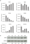
(A) qPCR of Wnt3, Wnt4, Wnt5a, Wnt5b, Wnt7a, and Wnt10b, in the mouse cerebellum from postnatal day 5 (P5) to adult. Wnt levels were normalized against β-2-microglobulin (B2M), hyporanthine-guanine phosphoribosyltransferase (HPRT1) and ribosomal subunit 18s (M18s). (B) WNT3 protein levels during cerebellar development. Immunoblotting of cerebellar lysates from P0 to P56 (adult) with an anti-WNT3 antibody showed an increase in WNT3 protein during cerebellar development.
Figure 2
Figure 2. WNT3 decreases GCP proliferation by inhibiting SHH dependent gene transcription.
(A) WNT3 decreased proliferation of GCPs by 40.3± 5.0% of the control (Con) (n=3) as measured by [3H]-Thymidine incorporation assay. (B) WNT3 did not increase cell death of GCPs in a TUNEL assay at 2 DIV (Con, 12.5±1.6%; WNT3, 13.1±0.7% (n=3)). (C) WNT3 decreased SHH dependent proliferation of GCPs (Con & SHH, 354.8±18.7%; WNT3 & SHH, 232.9±28.7% (n=3)). (D) WNT3 decreased Gli1, and Ptch1 mRNA levels after 6 h of treatment (Con=1; WNT3: Gli1=0.56±0.10, Gli2=0.90±0.10, Mycn= 0.99±0.05, Ptch1=0.58±0.09, and Ccnd1=0.89±0.09). (E) Treatment of GCPs with WNT3 antagonized the transcription of SHH targets. SHH was added for 24 h and WNT3 was added for 6h. (SHH/Con=1; SHH/WNT3: Gli1=0.62±0.04, Gli2=0.75±0.02, Mycn=1.12±0.05, Ptch1=0.70±0.11 and Ccnd1 =1.03±0.12.). (F) After 24 h of GLI1 overexpression in GCPs, Gli2, and Ptch1 mRNA levels increase in the control condition (Gli1=464±119, Gli2=2.04±0.25, Ptch1=1.59±0.14) and this effect is inhibited in the presence of WNT3 (Gli1=314±166, Gli2=1.14±0.3, Ptch1=0.71±0.12). Data represent the mean ± s.e.m.: *p<0.05, **p<0.01, ***p<0.001. NS, not significant.
Figure 4
Figure 4. GCP proliferation induced by WNT3 is not dependent on the BMP pathway.
(A) WNT3 and BMPs (BMP2, BMP4, BMP6, and BMP7) cooperated to decrease proliferation of GCPs as measured by [3H]-Thymidine incorporation assay. (B) The BMP inhibitor Noggin did not affect the WNT3-mediated decrease in proliferation. (WNT3, 59.9±4.6% of the control value; Con/Noggin, 93.5±1.7%; and WNT3/Noggin 46.4±2.9% (n=3).) (C) GCPs were stimulated for 4h at 1 DIV with WNT3 or BMP7 and analyzed by immunoblotting with the anti-phospho-SMAD1/5/8 antibody. The anti-SMAD1 antibody was used for normalization to obtain the percentage of phosphorylated SMAD1. (Con, 100±8.3%; WNT3, 133.8±33.8%; Con, 100±11.5%; and BMP7, 373.5±88.9% (n=3). Data represent the mean ± s.e.m.: *p<0.05, **p<0.01, ***p<0.001. NS, not significant.
Figure 3
Figure 3. WNT3 decreases the GCP proliferation marker Atoh1 and increases the GCP differentiation marker PAX6.
(A) WNT3 decreased Atoh1 mRNA levels after 6 h of treatment (Con=1; WNT3: Atoh1=0.52±0.06). (B) Treatment of GCPs with WNT3 decreased Atoh1 mRNA levels in the presence of SHH. SHH was added for 24 h and WNT3 was added for 6h. (SHH/Con=1; SHH/WNT3: Atoh1=0.47±0.04). (C) Overexpression of ATOH1 by retroviral infection of GCPs abrogated the WNT3 effect as measured by [3H]-Thymidine incorporation assay. GCPs were infected with retroviruses produced using pMSCV-GFP and pMSCV-Atoh1-GFP on day 1. WNT3 was added to the GCPs on day 2. (GFP/Con, 100±2.5%; GFP/WNT3, 77.4±3.3%; ATOH1-GFP/Con, 98.9±2.3%; ATOH1-GFP/WNT3, 92.6±3.8% (n=3).) (D) WNT3 decreased the mRNA levels of additional mitotic markers Ki67 and Notch2 after 24 h of treatment (Con=1; WNT3: Atoh1=0.71±0.07, Ki67=0.70±0.08, and Notch2=0.70±0.06). (E) P7 cerebellar slices were incubated with WNT3 for 24 h, and cryostat sections were immunostained with anti-PAX6 antibody, a marker for GCP differentiation. In control cultures (top panel), low levels of immunostaining are seen with anti-PAX6 antibody. WNT3 treatment (lower panel) increases the intensity of anti-PAX6 immunostaining in GCPs in the external granule layer (EGL), in postmitotic GCPs migrating across the molecular layer (ML, arrow) and in granule neurons in the internal granular layer (IGL) undergoing terminal differentiation. (F) WNT3 increased the mRNA levels of additional post-mitotic markers Zic2 and Gabra6 after 24 h of treatment (Con=1; WNT3: Zic2=3.22±0.54 and Gabra6=24.40±2.08). Data represent the mean ± s.e.m.: *p<0.05, **p<0.01, ***p<0.001. NS, not significant. Scale bar 100 µm.
Figure 5
Figure 5. WNT3 inhibits murine medulloblastoma formation.
(A) (Left panels) GCP-like tumor cells treated with WNT3 for 3 DIV. (Right panel) Quantitation of total cell number. (Day 0, Con and WNT3, 3.5x10⁵ cells; Day 3, Con, 7.7±0.3x10⁵ cells and WNT3, 3.8±0.3x10⁵ cells (n=5).) Scale bar 50 µm. (B) Cell cycle analysis by FACS of GCP-like tumor cells treated with WNT3 for 72 h. (Ink4c-/-Ptc1+/- tumor, Con, 11±1.4% and WNT3, 6±0% (n=2) and Ink4 c-/- p53 Fl/- Nestin-cre + tumor, Con, 24.5±2.12%, and WNT3, 12.5±2.12% (n=2).) After treatment with WNT3, no increase in apoptosis was observed using Annexin V (data not shown). (C) (Right image panels) Image of allografts established from medulloblastoma cells of a mouse (Ink4c-/-p53Fl/-Nestin-cre+) infected with control virus expressing YFP only or with virus expressing WNT3 and YFP. (Top right graph) Weight of allografts derived from cells infected with control YFP (0.82±0.16 g) or with WNT3 and YFP (0.30±0.13 g) (n=4). (Bottom right graph) Analysis by FACS of YFP expression in allografts derived from cells infected with control YFP (19.7±4.1%) or with WNT3 and YFP (2.9±1.2%) (n=3). (D) Tumors from athymic mice bearing allograft from FACS-sorted YFP-positive tumor cells infected with control virus (top panel, YFP) or WNT3 (bottom panel, WNT3-YFP), (n=2). (E) ATOH1 protein was decreased in WNT3 treated GCP-like tumor cells. Cyclopamine was used as a postitive control. β-actin was used as a loading control. Data represent the mean ± s.e.m.: *p<0.05, **p<0.01, ***p<0.001. NS, not significant.
Figure 6
Figure 6. WNT3 signaling is not mediated by β-catenin signaling.
(A, B,) GCPs were treated with WNT3 for 24 h and lysates analyzed by Western blot analysis. (A) Immunoblotting of GCP lysates with anti-active-β-catenin (Ac-β-cat) antibody showed that WNT3 did not induce β-catenin activation. The GSK-3 inhibitor BIO was used as positive control to increase activated β-catenin and activated β-catenin levels were compared to total β-catenin levels, using anti-β-catenin antibody (n=3). (B) Immunoblotting using anti-Phospho(P)-216-GSK-3β, anti-GSK-3β and anti-MYCN antibodies showed that WNT3 does not regulate β-catenin signaling. GAPDH was used as loading control. (C) Using a Luciferase assay, WNT3 fails to activate β-catenin signaling, which is reported by the expression of pTOPflash luciferase driven by the TCF/LEF promoter. LiCl was used as a positive control to activate β-catenin signaling. Data represent the mean ± s.e.m.: *p<0.05, **p<0.01, ***p<0.001. NS, not significant.
Figure 7
Figure 7. WNT3 activates the MAPK/ERK1,2 and ERK5 signaling pathway(s) in GCPs.
(A,B) GCPs were treated with WNT3 for 24 h and lysates analyzed by Western blot analysis. (A) WNT3 increases ERK1/2 and ERK5 phosphorylation in GCPs. Protein extracts were analyzed by immunoblotting with anti-P-ERK1/2, anti-ERK1/2, anti-P-ERK5, and anti-ERK5 antibodies (n=3). GAPDH was used as loading control. (B) Immunoblotting using anti-P-p38, anti-p38, anti-P-JNK and anti-JNK antibodies showed that WNT3 did not alter p38 or JNK activity. GAPDH was used as loading control. (C) Test of WNT3 specificity using a SRE Luciferase assay in GCPs. BDNF was used as a positive control regulator of MAPK signaling (n=3). (D) MAPK activity induced by WNT3 in GCPs is inhibited by the MEK inhibitor PD98059. SRE-luciferase assay in GCPs treated with control or WNT3, in the absence or presence of PD98059 (n=3). (E). The MEK inhibitor PD98059 reverses WNT3 inhibition of GCP proliferation. As measured by [3H]-Thymidine incorporation, PD98059 increases GCP proliferation by 15%±7%, WNT3 inhibits GCPs proliferation by 34±12% and PD98059 + WNT3 inhibits GCP proliferation by 7%±5% of the control (Con) (n=3). Data represent the mean ± s.e.m.: *p<0.05, **p<0.01, ***p<0.001. NS, not significant.
Figure 8
Figure 8. WNT3 induces activation of ERK1/2 ex vivo in P7 cerebellar slices and in medulloblastoma cells.
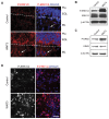
(A) P7 cerebellar slices were incubated with WNT3 for 24 h, and cryostat sections were immunostained with anti-P-ERK1/2 antibodies. Nuclei were stained with DRAQ5. P-ERK1/2 was induced in the external granule layer (EGL), and localized in the cytoplasm of the GCPs in the lower aspect of the EGL, where GCPs commence differentiation. P-ERK1/2 was also located in the molecular layer (ML) where postmitotic GCPs migrate toward the internal granule layer (IGL). Arrows point to an individual cell in the ML to highlight the cytoplasmic localization of P-ERK1/2. Scale bar 20 µm. (B C, D) GCP-like tumor cells were treated 24 h with WNT3. After treatment, GCP-like tumors were lysed and analysed by immunoblotting using (B) anti-P-ERK1/2 and anti-ERK1/2 antibodies or (C) anti-P-ERK5 and anti-ERK5 antibodies. β-ACTIN was used as loading control. (D) GCP-like tumor cells were immunostained after treatment using anti-P-ERK1/2 antibodies. Nuclei were stained with DRAQ5. WNT3 treatment increased the number of GCP-like tumor cells containing cytoplasmic P-ERK1/2, compared to the control. Scale bar 20 µm.
Figure 9
Figure 9. Interactions between WNT3 and SHH signaling pathways in GCPs.
In Hedgehog (Hh) signaling, Sonic Hedgehog (SHH) binds Patched (PTCH) receptor, relieving its inhibition of Smoothened (SMO), and stimulating GCP proliferation. In WNT3 signaling, WNT3 binds an undefined receptor to activate MAPK signaling via a non canonical WNT pathway to promote GCP differentiation, whereas other WNTs bind Frizzled (FRZ) receptor to activate the canonical β-catenin pathway and stimulate proliferation. We propose a model whereby regulation of MAPK activity by WNT3 signaling alters the balance between proliferation and differentiation in GCPs, decreasing proliferation and increasing differentiation.

References

    1. Wechsler-Reya RJ, Scott MP (1999) Control of neuronal precursor proliferation in the cerebellum by Sonic Hedgehog. Neuron 22: 103-114. doi:10.1016/S0896-6273(00)80682-0. PubMed: 10027293. - DOI - PubMed
    1. Chidambaram A, Goldstein AM, Gailani MR, Gerrard B, Bale SJ et al. (1996) Mutations in the human homologue of the Drosophila patched gene in Caucasian and African-American nevoid basal cell carcinoma syndrome patients. Cancer Res 56: 4599-4601. PubMed: 8840969. - PubMed
    1. Gilbertson RJ, Ellison DW (2008) The origins of medulloblastoma subtypes. Annu Rev Pathol 3: 341-365. doi:10.1146/annurev.pathmechdis.3.121806.151518. PubMed: 18039127. - DOI - PubMed
    1. Kool M, Koster J, Bunt J, Hasselt NE, Lakeman A et al. (2008) Integrated genomics identifies five medulloblastoma subtypes with distinct genetic profiles, pathway signatures and clinicopathological features. PLOS ONE 3: e3088. doi:10.1371/journal.pone.0003088. PubMed: 18769486. - DOI - PMC - PubMed
    1. Marino S (2005) Medulloblastoma: developmental mechanisms out of control. Trends Mol Med 11: 17-22. doi:10.1016/j.molmed.2004.11.008. PubMed: 15649818. - DOI - PubMed

Publication types

MeSH terms

Substances

LinkOut - more resources