. 2013 Jul;1(2):e0005.
doi: 10.1002/phy2.5. Epub 2013 Jun 26.

Molecular basis for impaired collateral artery growth in the spontaneously hypertensive rat: insight from microarray analysis

Affiliations

Molecular basis for impaired collateral artery growth in the spontaneously hypertensive rat: insight from microarray analysis

Joseph L Unthank et al. Physiol Rep. 2013 Jul.

Abstract

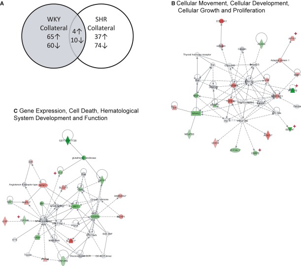
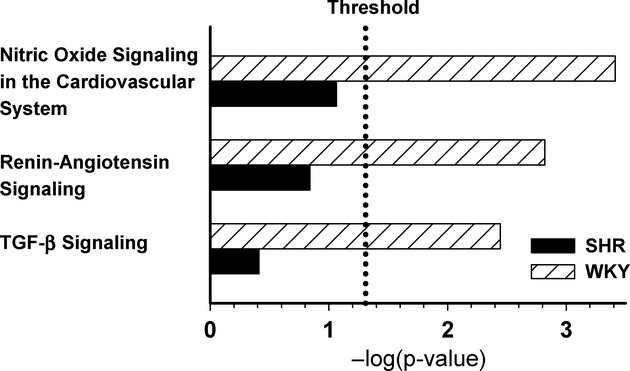
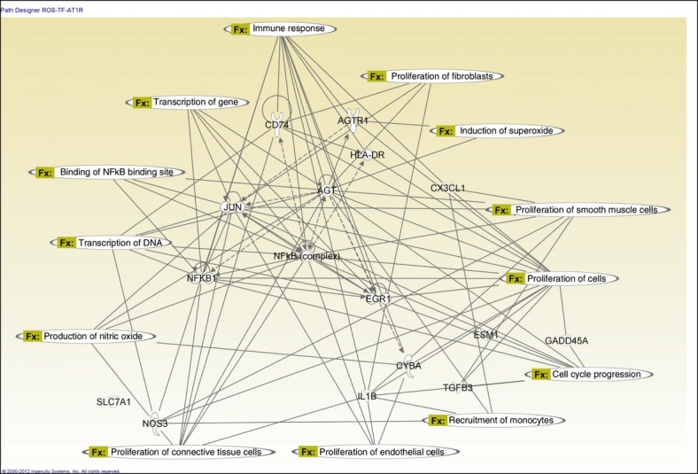
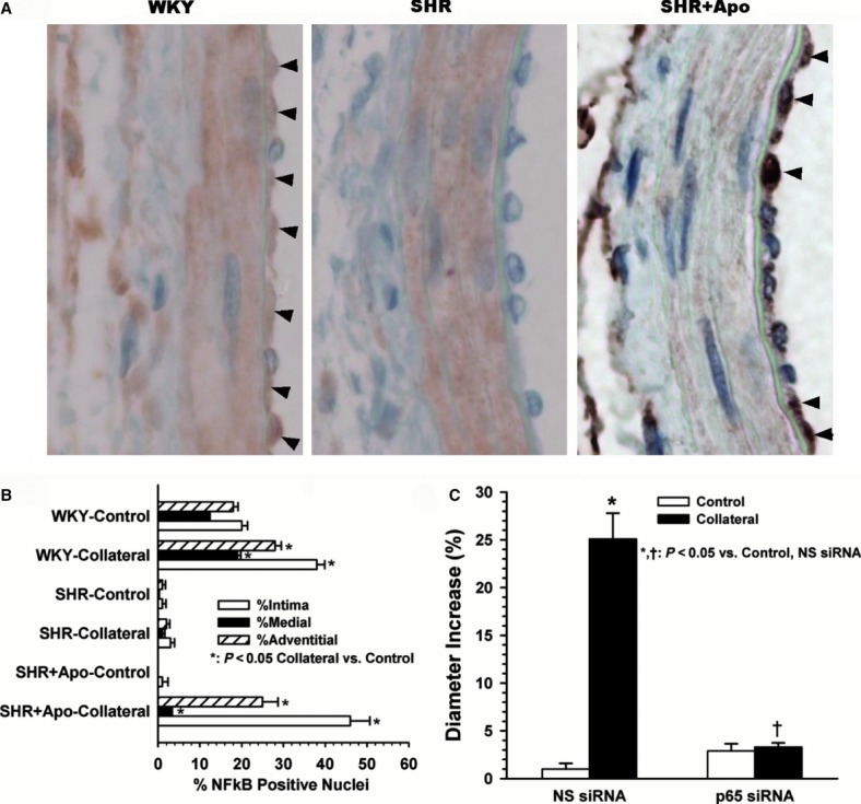
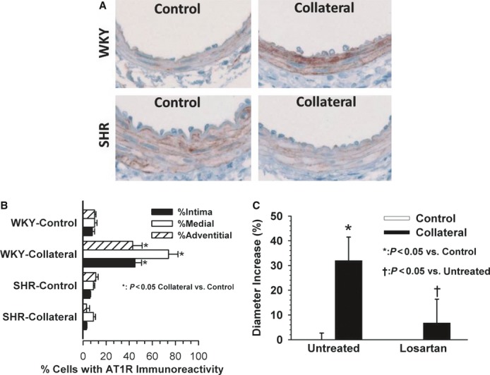
Analysis of global gene expression in mesenteric control and collateral arteries was used to investigate potential molecules, pathways, and mechanisms responsible for impaired collateral growth in the Spontaneously Hypertensive Rat (SHR). A fundamental difference was observed in overall gene expression pattern in SHR versus Wistar Kyoto (WKY) collaterals; only 6% of genes altered in collaterals were similar between rat strains. Ingenuity® Pathway Analysis (IPA) identified major differences between WKY and SHR in networks and biological functions related to cell growth and proliferation and gene expression. In SHR control arteries, several mechano-sensitive and redox-dependent transcription regulators were downregulated including JUN (-5.2×, P = 0.02), EGR1 (-4.1×, P = 0.01), and NFĸB1 (-1.95×, P = 0.04). Predicted binding sites for NFĸB and AP-1 were present in genes altered in WKY but not SHR collaterals. Immunostaining showed increased NFĸB nuclear translocation in collateral arteries of WKY and apocynin-treated SHR, but not in untreated SHR. siRNA for the p65 subunit suppressed collateral growth in WKY, confirming a functional role of NFkB. Canonical pathways identified by IPA in WKY but not SHR included nitric oxide and renin-angiotensin system signaling. The angiotensin type 1 receptor (AGTR1) exhibited upregulation in WKY collaterals, but downregulation in SHR; pharmacological blockade of AGTR1 with losartan prevented collateral luminal expansion in WKY. Together, these results suggest that collateral growth impairment results from an abnormality in a fundamental regulatory mechanism that occurs at a level between signal transduction and gene transcription and implicate redox-dependent modulation of mechano-sensitive transcription factors such as NFĸB as a potential mechanism.

Keywords: Arteriogenesis; collateral gene expression; microarray analysis; peripheral vascular disease.


Figures

Figure 1
Figure 1
Experimental design for microarray analysis. Microarray analysis utilizing the Affymetrix GeneChip Rat Genome U34A Array was performed on control and collateral mesenteric arteries of WKY and SHR (N = 4 each). Data were extracted using Affymetrix Microarray Suite 5.0 software and statistical analysis for a within subject design was performed after logarithmetic normalization. Data mining was then performed with IPA to identify molecules with the greatest expression changes, top biological functions, and canonical pathways. Motif Modeler was used to find predicted transcription factor binding sites in molecules with altered expression in collateral relative to control arteries.
Figure 2
Figure 2
Fundamental differences in collateral gene expression. (A) Venn diagram of number of genes with increased (↑) or decreased (↓) expression in collateral artery relative to same animal control artery for WKY and SHR (P ≤ 0.05, fold change ≥1.25×). An IPA comparison analysis of the molecules with altered expression (125 in WKY, 111 in SHR) indicates 222 genes unique to either set, with only 14 common to both sets and demonstrating similar expression changes. The upregulated genes with common expression between WKY and SHR included AP1S1 (adaptor-related protein complex 1, sigma 1 subunit), C6orf115 (ABRA C-terminal like), LRRC59 (leucine rich repeat containing 59), and PTPN2 (protein tyrosine phosphatase, nonreceptor type 2); those downregulated were ATP1A2 (ATPase, Na+/K+ transporting, alpha 2 polypeptide), COX8A (cytochrome c oxidase subunit VIIIA (ubiquitous)), CST3 (cystatin C), ECH1 (enoyl CoA hydratase 1, peroxisomal), FUCA1 (fucosidase, alpha-L- 1, tissue), IDH2 (isocitrate dehydrogenase 2 (NADP+), mitochondrial), LDHB (lactate dehydrogenase B), PPA2 (pyrophosphatase (inorganic) 2), PPP1R1A (protein phosphatase 1, regulatory (inhibitor) subunit 1A), and ZFP36L1(zinc finger protein 36, C3H type-like 1). The two IPA highest scored networks in WKY were Cellular Movement, Cellular Development, Cellular Growth and Proliferation shown in (B), and Gene Expression, Cell Death, Hematological System Development and Function depicted in (C). Molecules with significant up- and downregulation are identified with red and green shading, respectively. Comparison of these molecules between WKY and SHR demonstrated fundamental differences. For those genes in the Cellular Movement, Cellular Development, Cellular Growth and Proliferation, only AP1S1(adaptor-related protein complex 1, sigma 1 subunit), ATP1A2, EEF2K (eukaryotic elongation factor-2 kinase), and CSRP2 (cysteine and glycine-rich protein 2) had altered expression in SHR (red +). Similarly, within the Gene Expression, Cell Death, Hematological System Development and Function network, only CLU (clusterin) and FUCA (fucosidase, alpha-L- 1, tissue) had altered collateral expression in SHR (red +). □ = cytokine, ⋄ = enzyme, ○ = other, horizontal oval = transcription regulator, ▲= phosphatase, ▼ = kinase, vertical oval = transmembrane receptor, trapezoid = transporter. Lines without arrows indicate binding, lines with arrows indicate stimulation, solid lines indicate direct interaction, and dashed lines indirect.
Figure 3
Figure 3
Canonical pathways within IPA known to the authors to have important roles in various types of arterial remodeling and which exhibited significant alterations in WKY but not SHR included signaling pathways associated with nitric oxide, the renin–angiotensin system (RAS), and transforming growth factor-beta as shown in this figure.
Figure 4
Figure 4
IPA constructed network of key molecules selected from those with the greatest fold changes (Esm1, CX3CL1, CD74, HLA-DR, TGFβ3, ILβ1, GADD45α, SLC7A1), members of highest scored Canonical pathways (eNOS, SLC7A1, AGT, AT1R, TGFβ3), and the NAD(P)H oxidase component, CYBA. Altered regulation of several of these molecules was confirmed by RT-PCR (Table 8). IPA generated connections between the molecules and overlaid specific functions.
Figure 5
Figure 5
Nuclear localization of NF-κB during successful and impaired collateral growth. (A) Representative images showing NF-κB immunoreactivity (brown) in collateral arteries 3 days after arterial ligation in WKY, SHR, and apocynin pretreated SHR (SHR+Apo). Nuclear localization is apparent especially within the intima of WKY and SHR+Apo, as indicated by arrows, but not in SHR. (B) Analysis of the percentage of cells with immunoreactivity in each wall layer indicates a statistical increase in all wall layers of collaterals from WKY and SHR+Apo relative to same animal controls, (n ≥ 3, *P ≤ 0.001). (C) Inhibition of p65 expression suppressed collateral growth. Paired comparisons of control and collateral arteries before and 7 days after arterial ligation demonstrated significant collateral enlargement in WKY administered a control nonsense siRNA (*P ≤ 0.001, n = 4) but not in WKY pretreated with siRNA to p65 (P ≤ 0.001 vs. nonsense siRNA collateral, n = 3). No effect of p65 siRNA was observed on the diameters of control arteries.
Figure 6
Figure 6
Potential role of the AGTR1 in collateral growth. (A) Representative images showing AGTR1 immunoreactivity (brown) in WKY and SHR control and collateral arteries 3 days after arterial ligation. Relative to same animal controls, a remarkable increase is observed in all wall layers of the WKY but not SHR collateral. (B) Analysis of the percentage of cells with immunoreactivity in each wall layer indicates a statistical increase in all wall layers of the WKY collateral and a decrease in the adventitia of the SHR collateral compared to same animal control artery (n = 3, *P ≤ 0.05). C. Effect of AGTR1 blockade by losartan on collateral diameter enlargement in WKY. Paired comparisons of control and collateral arteries before and 7 days after arterial ligation demonstrated significant collateral enlargement in WKY-untreated animals (*P ≤ 0.001) but not in WKY pretreated with losartan (P = 0.215), which prevents collateral growth in WKY. No effect of losartan was observed on the diameters of control arteries, consequently the open bars are not apparent on the graph (n = 5). The combined data suggest that the upregulation and activation of the AGTR1 is not only correlated with, but is also required for collateral growth in the young normotensive WKY.

References

    1. Ahimastos AA, Lawler A, Reid CM, Blombery PA, Kingwell BA. Brief communication: ramipril markedly improves walking ability in patients with peripheral arterial disease: a randomized trial. Ann. Intern. Med. 2006;144:660–664. - PubMed
    1. Aitkenhead M, Wang SJ, Nakatsu MN, Mestas J, Heard C, Hughes CC. Identification of endothelial cell genes expressed in an in vitro model of angiogenesis: induction of ESM-1, (beta)ig-h3, and NrCAM. Microvasc. Res. 2002;63:159–171. - PubMed
    1. Allen RG, Tresini M. Oxidative stress and gene regulation. Free Radic. Biol. Med. 2000;28:463–499. - PubMed
    1. Bechard D, Scherpereel A, Hammad H, Gentina T, Tsicopoulos A, Aumercier M, et al. Human endothelial-cell specific molecule-1 binds directly to the integrin CD11a/CD18 (LFA-1) and blocks binding to intercellular adhesion molecule-1. J. Immunol. 2001;167:3099–3106. - PubMed
    1. Béchard D, Gentina T, Delehedde M, Scherpereel A, Lyon M, Aumercier M, et al. Endocan is a novel chondroitin sulfate/dermatan sulfate proteoglycan that promotes hepatocyte growth factor/scatter factor mitogenic activity. J. Biol. Chem. 2001;276:48341–48349. - PubMed

LinkOut - more resources