. 2013 Jul;5(3):126-9.

Stabilization of the Central Part of Tropomyosin Molecule Alters the Ca2+-sensitivity of Actin-Myosin Interaction

Affiliations

Stabilization of the Central Part of Tropomyosin Molecule Alters the Ca2+-sensitivity of Actin-Myosin Interaction

D V Shchepkin et al. Acta Naturae. 2013 Jul.

Abstract

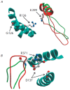
We show that the mutations D137L and G126R, which stabilize the central part of the tropomyosin (Tm) molecule, increase both the maximal sliding velocity of the regulated actin filaments in the in vitro motility assay at high Са(2+) concentrations and the Са(2+)-sensitivity of the actin-myosin interaction underlying this sliding. Based on an analysis of the recently published data on the structure of the actin-Tm-myosin complex, we suppose that the physiological effects of these mutations in Tm can be accounted for by their influence on the interactions between the central part of Tm and certain sites of the myosin head.

Keywords: actin-myosin interaction; in vitro motility assay; regulation of muscle contraction; tropomyosin.


Figures

Fig. 1
Fig. 1
The average sliding velocity of the thin filaments along the myosin-coated surface as a function of the Ca2+ concentration. A: average data for 2–3 experiments with each of the Tm mutants. Vertical lines show the standard deviations. B: the same data as in A normalized for the maximum velocity
Fig. 2
Fig. 2
The structural model of the contact area of a myosin head strongly bound to actin with Tm on the surface of the thin filament. Only some parts of the myosin head adjacent to the amino acid residues 126 (A) and 137 (B) in the central part of Tm are shown. Actin and other parts of myosin and Tm are not shown. The model was obtained from the structure of the actin–myosin–Tm complex ([8], pdb code 4A7H) by superimposing the upper 50-kDa domain of the myosin head of chicken fast skeletal muscle myosin II (pdb code 2MYS) instead of the same domain of myosin-I used in [8]. Segments of the Tm double α-helix are shown by blue ribbons, the parts of the myosin-I head used in [8] are shown in red, and those of the head of skeletal muscle myosin-II are green. The residue R126 of the Tm G126R mutant (A) and a ‘non-canonical’ Tm residue D137 (B), which was replaced with Leu in the Tm mutant D137L/C190A, as well as the charged myosin residues K399 (A) and R371 (B) located in close proximity to these Tm residues are shown in a ‘ball-and-stick’ atomic representation. The distance between the charged atoms of myosin residue K399 and Tm residue R126 (A) in the model was 8.8 A°, and that between myosin R371 and Tm D137 (B) was 4.7 A°. The model and the picture were prepared using ICM-Browser (MolSoft, CA, USA)

References

    1. McKillop D.F.A., Geeves M.A.. Biophys. J. 1993;65:693–701. - PMC - PubMed
    1. Nevzorov I.A., Levitsky D.I.. Biochemistry (Moscow). 2077;76(13):1507–1527. - PubMed
    1. Sumida J.P., Wu E., Lehrer S.S.. J. Biol. Chem. 2008;283:6728–6734. - PubMed
    1. Nevzorov I.A., Nikolaeva O.P., Kainov Y.A., Redwood C.S., Levitsky D.I.. J. Biol. Chem. 2011;286:15766–15772. - PMC - PubMed
    1. Shchepkin D.V., Kopylova G.V., Nikitina L.V., Katsnelson L.B., Bershitsky S.Y.. Biochem. Biophys. Res. Commun. 2010;401(1):159–163. - PubMed

LinkOut - more resources