

Fibromyxoma of the lateral skull base in a child: case report
- PMID: 24303345
- PMCID: PMC3836972
- DOI: 10.1055/s-0033-1351115
Fibromyxoma of the lateral skull base in a child: case report
Abstract
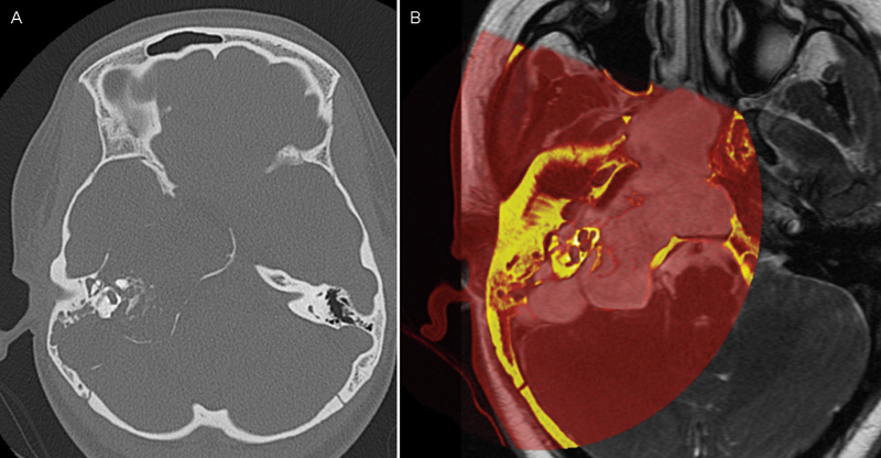
Purpose Fibromyxomas and myxomas are benign tumors of mesenchymal origin usually found outside the nervous system, most commonly in the atrium of the heart. They can also arise in the mandible or maxilla, but it is exceedingly rare to find them within the skull base. The history, histologic features, and the literature, with emphasis on other pediatric cases, are reviewed for this uncommon skull base neoplasm. Methods We describe the case of a 13-year-old girl who presented with a 1-year history of facial weakness, numbness, and hearing loss. A large locally destructive tumor centered in the petrous bone was found on magnetic resonance imaging. Results A mastoidectomy combined with a middle fossa craniotomy was performed for gross total resection. The child is disease free 12 months after surgery. Conclusion Diagnosis could not be made solely on radiographic studies because of the lack of pathognomonic imaging features. Radical resection provided the patient the best chance of cure. Long-term surveillance is necessary to monitor for tumor recurrence.
Keywords: benign; child; fibromyxoma; mesenchymal; myxoma; pediatric; petrous; skull base; tumor.
Figures


References
-
- Chapman P R Shah R Curé J K Bag A K Petrous apex lesions: pictorial review AJR Am J Roentgenol 2011196(3, Suppl):WS26–WS37.; quiz S40–S43 - PubMed
-
- Erdem Y, Koktekir E, Bayar M A, Yilmaz A, Caydere M. Characterization of an intracranial neurothekeoma: case report. Turk Neurosurg. 2012;22(1):109–112. - PubMed
-
- Frank E, Deruaz J P, de Tribolet N. Chondromyxoid fibroma of the petrous-sphenoid junction. Surg Neurol. 1987;27(2):182–186. - PubMed
-
- Oruckaptan H H, Sarac S, Gedikoglu G. Primary intracranial myxoma of the lateral skull base: a rare entity in clinical practice. Turk Neurosurg. 2010;20(1):86–89. - PubMed
-
- Osterdock R J Greene S Mascott C R Amedee R Crawford B E Primary myxoma of the temporal bone in a 17-year-old boy: case report Neurosurgery 2001484945–947.; discussion 947–948 - PubMed
LinkOut - more resources
Full Text Sources
Other Literature Sources