. 2014 Feb;91(3):618-34.
doi: 10.1111/mmi.12482. Epub 2014 Jan 7.

The multifunctional role of the pallilysin-associated Treponema pallidum protein, Tp0750, in promoting fibrinolysis and extracellular matrix component degradation

Affiliations

The multifunctional role of the pallilysin-associated Treponema pallidum protein, Tp0750, in promoting fibrinolysis and extracellular matrix component degradation

Simon Houston et al. Mol Microbiol. 2014 Feb.

Abstract

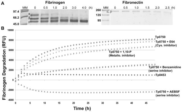
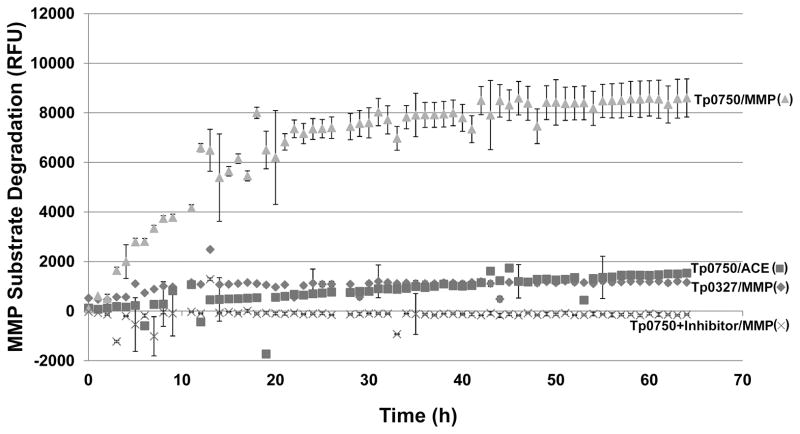
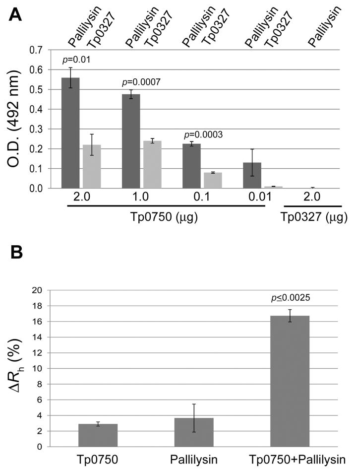
The mechanisms that facilitate dissemination of the highly invasive spirochaete, Treponema pallidum, are incompletely understood. Previous studies showed the treponemal metalloprotease pallilysin (Tp0751) possesses fibrin clot degradation capability, suggesting a role in treponemal dissemination. In the current study we report characterization of the functionally linked protein Tp0750. Structural modelling predicts Tp0750 contains a von Willebrand factor type A (vWFA) domain, a protein-protein interaction domain commonly observed in extracellular matrix (ECM)-binding proteins. We report Tp0750 is a serine protease that degrades the major clot components fibrinogen and fibronectin. We also demonstrate Tp0750 cleaves a matrix metalloprotease (MMP) peptide substrate that is targeted by several MMPs, enzymes central to ECM remodelling. Through proteomic analyses we show Tp0750 binds the endothelial fibrinolytic receptor, annexin A2, in a specific and dose-dependent manner. These results suggest Tp0750 constitutes a multifunctional protein that is able to (1) degrade infection-limiting clots by both inhibiting clot formation through degradation of host coagulation cascade proteins and promoting clot dissolution by complexing with host proteins involved in the fibrinolytic cascade and (2) facilitate ECM degradation via MMP-like proteolysis of host components. We propose that through these activities Tp0750 functions in concert with pallilysin to enable T. pallidum dissemination.


Figures

Fig. 1
Fig. 1. tp0750 and tp0751 comprise a two-gene operon
A. The potential for tp0750 and tp0751 to comprise a two-gene operon was investigated by analysis of the T. pallidum genome sequence using the Artemis Genome Browser. FS, forward strand; RS, reverse strand; BN, base pair number; FRF1-3, forward reading frames 1–3; RFR1-3, reverse reading frames 1–3. Arrows indicate the location and orientation of gene-specific primers used in the RT-PCR reaction. B. The two-gene operon organization of tp0750 and tp0751 was confirmed by analysing T. pallidum RNA by RT-PCR. The cDNA product amplified from the tp0750/tp0751 primer pair (RT+) and the PCR product of the same size amplified from T. pallidum genomic DNA (gDNA) are shown. A cDNA product was not detected when reverse transcriptase was omitted from the RT-PCR reaction (RT−).
Fig. 2
Fig. 2. Tp0750 molecular modelling predicts similarity to a MIDAS- and vWFA domain-containing protein
A. Schematic representation of the results of SignalP and NCBI conserved domain database analyses performed on the Tp0750 amino acid sequence. Indicated are the predicted SpI (signal peptidase I) cleavage site (between A22 and G23, arrow) and Metal Ion-Dependent Adhesion Site (MIDAS)-containing vWFA (von Willebrand Factor type A) domain (V29-T147, grey shading; rectangles indicate MIDAS site residues). B. Model of Tp0750 generated by the Modeller program predicts a central 5-stranded beta sheet of mixed parallel and anti-parallel beta strands and 5 framing alpha helices (upper panel). The predicted structure of Tp0750 was modelled using integrin α2β1 (PDB 1DZI), which contains both a vWFA domain and a MIDAS motif (lower panel). Right (inset) panels: residues comprising the MIDAS motif within Tp0750 (upper panel) are conserved in sequence and structure with the cobalt-binding MIDAS motif of integrin α2β1 (lower panel), suggesting Tp0750 has metal ion-binding capability.
Fig. 3
Fig. 3. Tp0750 exhibits specific binding to annexin A2
A. Binding assays were performed to compare the attachment of recombinant Tp0750 (2 μg) and a negative control protein (Tp0453; 2 μg) to immobilized annexin A2 (0.5 μg) and syntaxin-2 (0.5 μg). Tp0750 exhibited a statistically significant level of binding to annexin A2 (p≤0.0072) when compared to the binding of the negative control to annexin A2 and Tp0750 to syntaxin-2. B. Dose-dependent binding assays confirmed the specificity of the Tp0750-annexin A2 interaction. Similar dose-dependent binding responses were observed upon use of (i) varying concentrations of immobilized annexin A2 (0.005–0.5 μg per well) and a constant concentration of soluble Tp0750 (2 μg per well) (left panel), and (ii) a constant concentration of immobilized annexin A2 (0.5 μg per well) and varying concentrations of soluble Tp0750 (0.01–2.0 μg per well) (right panel). Average readings from triplicate measurements are presented with bars indicating standard error (SE), and the results are representative of three independent experiments. For statistical analyses, the Student’s two-tailed t test was used to compare the attachment of (i) Tp0750 to annexin A2 and the control host protein syntaxin-2 and (ii) Tp0750 and the control recombinant protein Tp0453 to annexin A2.
Fig. 4
Fig. 4. Tp0750 binds human fibrinogen
Attachment of Tp0750 and a negative control protein (Tp0453; 2 μg) to immobilized fibrinogen and laminin (0.5 μg) was investigated. Tp0750 exhibited statistically significant levels of binding to fibrinogen (p=0.0005) when compared to the level of binding by the negative control. Average readings of triplicate wells are presented with bars indicating standard error and the results are representative of three independent experiments. For statistical analyses, attachment to the host proteins by Tp0750 was compared to attachment by Tp0453 using the Student’s two-tailed t test.
Fig. 5
Fig. 5. Tp0750 mediates degradation of fibrinogen and fibronectin and is inhibited by serine protease inhibitors
A. Protein degradation assays were performed to determine if Tp0750 is capable of degrading fibrinogen and fibronectin. Recombinant Tp0750_G23-A199 was incubated with human fibrinogen (20 μg Tp0750 and 100 μg fibrinogen) or fibronectin (2 μg Tp0750 and 30 μg fibronectin) for 4 h and 2.5 h, respectively. Samples (estimated to be 10 μg fibrinogen and 1.5 μg fibronectin in the absence of degradation) were removed at various time points and degradation of the fibrinogen α-, β-, and γ-chains (left panel) and co-migrating fibronectin α- and β-chains (right panel) was analyzed by SDS-PAGE. The negative control, Tp0453 (20 μg), failed to degrade fibrinogen or fibronectin (data not shown). Numbers to the left of the lanes indicate the size (kDa) of the corresponding molecular mass (MM) markers. B. Fluorescence-based degradation assays were performed to investigate the effect of protease inhibitors on the fibrinogenolytic activity of Tp0750. Recombinant proteins (Tp0453 and Tp0750_G23-A199) (1 μg) were pre-incubated for 1 h in the presence or absence of 1 mM 1,10 phenanthroline (1,10-P), 1 mM 4-(2-Aminoethyl) benzenesulfonyl fluoride hydrochloride (AEBSF), 1 mM benzamidine, or 200 μM trans-epoxysuccinyl-L-leucylamido (4-guanidino) butane (E64) prior to incubation with FITC-labelled fibrinogen (10 μg) for 48 h. The degree of fibrinogen degradation was measured every hour by detecting the increase in relative fluorescence units (RFU) using standard fluorescein excitation/emission filters. The increase in RFU for the two recombinant proteins +/− inhibitors is shown. Average fluorescence intensity readings from triplicate measurements are presented with bars indicating standard error (SE) and the results are representative of three independent experiments.
Fig. 6
Fig. 6. Tp0750-mediated MMP substrate degradation
A fluorescence-based degradation assay was used to determine if recombinant Tp0750 is capable of cleaving a MMP substrate. A negative control protein (Tp0327; 400 ng) and Tp0750 (400 ng) +/− the serine protease inhibitor AEBSF were incubated with either the MMP substrate (MMP; 2.5 μg) or the negative control substrate (ACE; 2.5 μg) (Tp0750 only) for 0–64 h. MMP substrate cleavage was measured by detecting the increase in relative fluorescence units (RFU) using standard fluorescein excitation/emission filters (320 nm/420 nm). Average fluorescence intensity readings from triplicate measurements are presented with bars indicating standard error (SE) and the results are representative of three independent experiments.
Fig. 7
Fig. 7. Interaction capability of Tp0750 and pallilysin
A. Dose-dependent binding assays were performed to determine the ability of His-tagged pallilysin (Tp0751_HAXXH mutant) (2 μg per well) and a His-tagged negative control protein (Tp0327) (2 μg per well) to bind varying concentrations of immobilized tagless Tp0750 (0.01 μg-2 μg per well). Pallilysin exhibited statistically significant levels of binding to immobilized Tp0750 (0.1–2 μg per well; p<0.01) when compared to the level of binding of the negative control (Tp0327) to immobilized Tp0750 and pallilysin to the immobilized tagless control protein (Tp0327). Tp0750 and the negative control protein Tp0327 exhibited similar coating efficiencies (data not shown). Average readings from triplicate measurements are presented with bars indicating standard error (SE) and the results are representative of three independent experiments. For statistical analyses, attachment capabilities were compared using the Student’s two-tailed t test. B. DLS analyses were performed to further confirm the Tp0750-pallilysin interaction. Tp0750 and pallilysin (0.9 mg ml−1) were incubated alone and together at room temperature for 60 min, centrifuged for 20 min and analyzed by DLS after total incubation times of 80 and 180 min. Average readings from triplicate measurements are presented as the percent difference between the hydrodynamic radius measurements recorded at 80 and 180 min (ΔRh [%]) with bars indicating standard error. The results are representative of three independent experiments. The Tp0750-pallilysin mixed sample exhibited a statistically significant higher increase in ΔRh when compared to Tp0750 or pallilysin alone (Student’s two-tailed t test: p≤0.0025).
Fig. 8
Fig. 8. Proposed model of the role of Tp0750/pallilysin in T. pallidum dissemination via the bloodstream
(1) Surface-exposed pallilysin ( formula image) mediates attachment of Treponema pallidum ( formula image
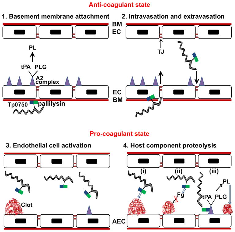
) to the laminin-rich basement membrane (BM) that underlies the endothelial cells (EC) of blood vessels. The unperturbed endothelium exists primarily in an anti-coagulant state due to the expression of profibrinolytic factors including the fibrinolytic complex comprising annexin A2 and S100A10 ( formula image) which binds tissue plasminogen activator (tPA) and plasminogen (PLG) resulting in plasmin (PL)-mediated clot dissolution; (2) T. pallidum intravasation and extravasation: pallilysin degrades the basement membrane. Tp0750 ( formula image), through interaction with annexin A2, localizes T. pallidum to areas of tight junction (TJ) remodeling; (3) Activation of endothelial cells occurs upon infection with T. pallidum leading to the formation of a pro-coagulant endothelial surface. On the surface of activated endothelial cells (AEC), the coagulation cascade is initiated resulting in the generation of vascular clots composed of fibrin, fibronectin, and platelets; (4) Tp0750/pallilysin-mediated host component proteolysis: (i) Direct clot degradation via pallilysin-mediated fibrinolysis and Tp0750-mediated fibronectinolysis, (ii) clot formation inhibition via Tp0750/pallilysin-mediated fibrinogen (Fg) degradation, and (iii) Tp0750-annexin A2 interaction localizes T. pallidum to the immediate vicinity of plasmin-mediated vascular fibrinolysis. Open arrows indicate direct proteolysis of host components. Closed arrows indicate an intermediate pro-protease activation step prior to direct proteolysis of host components.

References

    1. Abdallah AM, Verboom T, Hannes F, Safi M, Strong M, Eisenberg D, et al. A specific secretion system mediates PPE41 transport in pathogenic mycobacteria. Mol Microbiol. 2006;62:667–679. - PubMed
    1. Ait-Oufella H, Maury E, Lehoux S, Guidet B, Offenstadt G. The endothelium: physiological functions and role in microcirculatory failure during severe sepsis. Intensive Care Med. 2010;36:1286–1298. - PubMed
    1. Bharadwaj A, Bydoun M, Holloway R, Waisman D. Annexin A2 heterotetramer: structure and function. Int J Mol Sci. 2013;14:6259–6305. - PMC - PubMed
    1. Boucher DJ, Adler B, Boyce JD. The Pasteurella multocida nrfE gene is upregulated during infection and is essential for nitrite reduction but not for virulence. J Bacteriol. 2005;187:2278–2285. - PMC - PubMed
    1. Cameron CE. Identification of a Treponema pallidum laminin-binding protein. Infect Immun. 2003;71:2525–2533. - PMC - PubMed

Publication types

LinkOut - more resources