. 2014 Mar;39(5):771-87.
doi: 10.1111/ejn.12439. Epub 2013 Dec 5.

Connexin36 in gap junctions forming electrical synapses between motoneurons in sexually dimorphic motor nuclei in spinal cord of rat and mouse

Affiliations

Connexin36 in gap junctions forming electrical synapses between motoneurons in sexually dimorphic motor nuclei in spinal cord of rat and mouse

W Bautista et al. Eur J Neurosci. 2014 Mar.

Abstract

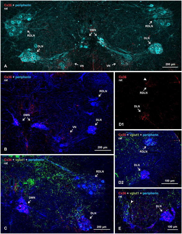
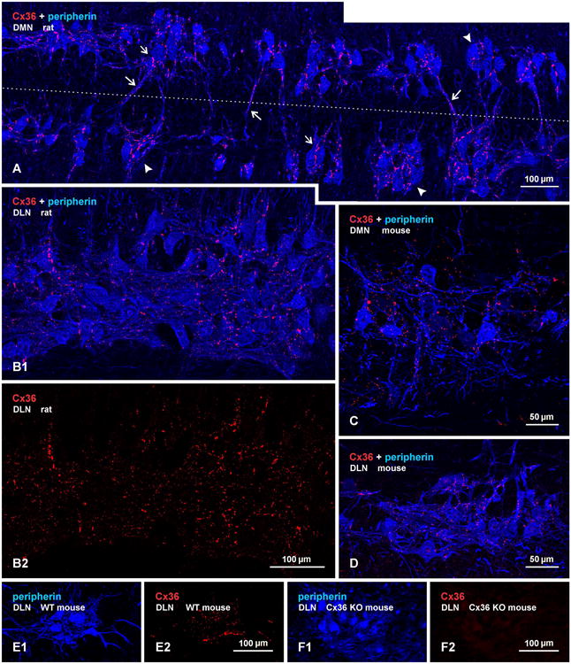
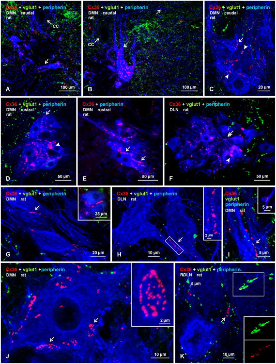
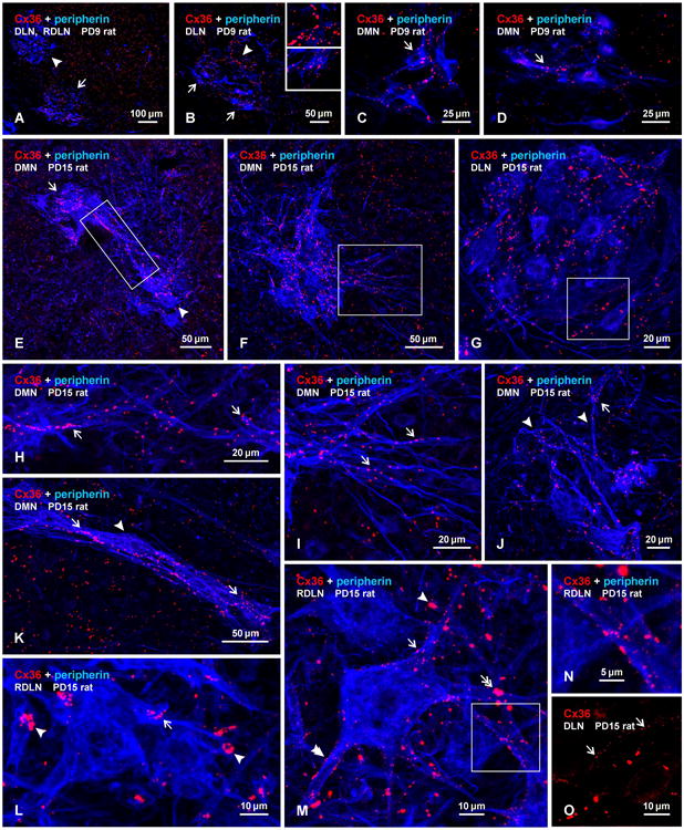
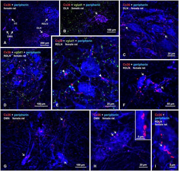
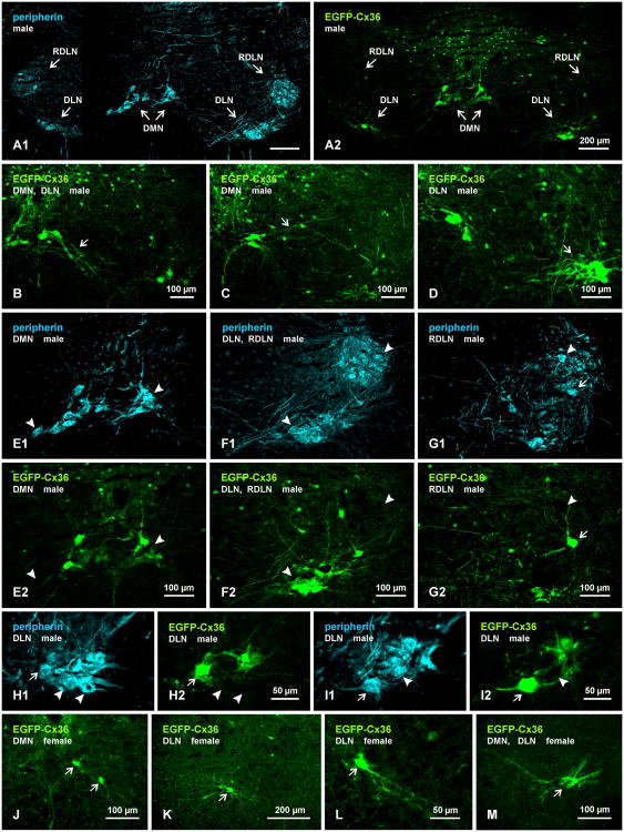
Pools of motoneurons in the lumbar spinal cord innervate the sexually dimorphic perineal musculature, and are themselves sexually dimorphic, showing differences in number and size between male and female rodents. In two of these pools, the dorsomedial nucleus (DMN) and the dorsolateral nucleus (DLN), dimorphic motoneurons are intermixed with non-dimorphic neurons innervating anal and external urethral sphincter muscles. As motoneurons in these nuclei are reportedly linked by gap junctions, we examined immunofluorescence labeling for the gap junction-forming protein connexin36 (Cx36) in male and female mice and rats. Fluorescent Cx36-labeled puncta occurred in distinctly greater amounts in the DMN and DLN of male rodents than in other spinal cord regions. These puncta were localized to motoneuron somata, proximal dendrites, and neuronal appositions, and were distributed either as isolated or large patches of puncta. In both rats and mice, Cx36-labeled puncta were associated with nearly all (> 94%) DMN and DLN motoneurons. The density of Cx36-labeled puncta increased dramatically from postnatal days 9 to 15, unlike the developmental decreases in these puncta observed in other central nervous system regions. In females, Cx36 labeling of puncta in the DLN was similar to that in males, but was sparse in the DMN. In enhanced green fluorescent protein (EGFP)-Cx36 transgenic mice, motoneurons in the DMN and DLN were intensely labeled for the EGFP reporter in males, but less so in females. The results indicate the presence of Cx36-containing gap junctions in the sexually dimorphic DMN and DLN of both male and female rodents, suggesting coupling of not only sexually dimorphic but also non-dimorphic motoneurons in these nuclei.

Keywords: EGFP-Cx36 reporter; gap junctions; immunofluorescence; motoneurons.


Figures

Fig. 1
Fig. 1
Overview of sexually dimorphic motor nuclei in relation to immunofluorescence labelling for Cx36 and vglut1 in transverse spinal cord sections of adult male rat. Motoneurons are labelled for peripherin, pseudo colored sky blue to better visualize motor nuclei (A), or deep blue to better visualize Cx36 and vglut1 (B-E). (A) Bilateral view at a lower lumbar level showing locations of, and labelling for Cx36 in, the DMN, DLN, RDLN and VN. (B-E) Images showing relative densities of labelling for Cx36 and/or vglut1, with dense labelling in the DMN and DLN (B-D) and moderate labelling in the VN (B). Sparse labelling is seen in the RDLN, as shown at a caudal level (C) and a more rostral level (D), with labelling for Cx36 shown alone (D1, arrowhead) and after overlay with labelling for peripherin and vglut1 (D2). Labelling for vglut1 is largely absent in the DMN and DLN (C,D2), and is of moderate density in the RDLN (C,D2). (E) Comparison of the absence of vglut1-terminals in DLN (arrow) vs. their presence in an adjacent unidentified motor nucleus (arrowhead).
Fig. 2
Fig. 2
Immunofluorescence labelling of Cx36 in association with peripherin-positive motoneurons in horizontal sections through the DMN and DLN of adult rat and mouse. (A) The DMN in rat, showing intermittent clusters of motoneurons (arrowheads), with some of these neurons having contralaterally projecting dendrites. Midline is shown by dotted line. Cx36-puncta are seen densely concentrated along appositions between motoneuronal somata in the clusters (small arrows) and along bundles of intermingled dendrites spanning the midline (large arrows). (B1,B2) The same field of the DLN in rat, showing overlay of labelling for peripherin and Cx36 (B1), where Cx36-puncta are seen associated with nearly all motoneurons, and labelling for Cx36 alone (B2), where a similar density of Cx36-puncta is seen in the medial (lower half of field) and lateral (upper half of field) portions of the nucleus containing ischiocavernosus and urethral sphincter motoneurons, respectively. (C,D) The DMN (C) and DLN (D) in adult mouse, showing in each nucleus a similarly large proportion of motoneurons invested with Cx36-puncta as seen in rat. (E,F) Images of the DLN from a wild-type mouse showing the same field labelled for peripherin and Cx36 (E1,E2), and from a Cx36 knockout mouse showing the same field with absence of Cx36 among peripherin-positive motoneurons (F1,F2).
Fig. 3
Fig. 3
Confocal triple immunofluorescence demonstrating patterns of Cx36-puncta and vglut1-terminals associated with motoneurons in DMN, DL, and RDLN in transverse sections of adult male rat spinal cord. (A,B) Images at a caudal level of the DMN, showing closely apposed peripherin-positive motoneurons, with Cx36-puncta lining dendrites directed either horizontally across the midline (A, arrow) or directed dorsally and laterally (B, arrows) in the vicinity of the central canal (cc). (C-F) Magnifications of clusters of motoneuron somata in the caudal DMN (C), rostral DMN (D,E) and DLN (F), showing punctate appearance of labelling for Cx36 and absence of diffuse intracellular labelling for Cx36. Also evident are both large heterogeneously sized patches of Cx36 immunofluorescence (C,D, arrowheads), fine dispersed Cx36-puncta (F, double arrows), and Cx36-puncta localized around the periphery of motoneurons (single arrows). (G-I) Images of the DMN, showing Cx36-puncta localized at soma-somatic (G, arrow; also shown in inset), dendro-somatic (H, arrow; boxed area magnified in inset), and dendro-dendritic (I, arrow; and inset) appositions between peripherin-positive motoneurons. (J) Magnification of a peripherin-positive motoneuron in the DMN, showing large patches of labelling for Cx36 (arrows), consisting of clusters of individual Cx36-puncta (one cluster shown in inset). (K) Magnification of the RDLN, showing sparse distribution of vgut1-terminals among motoneurons, and Cx36-puncta with (boxed area, shown separately as green and red labels in inset) and without (arrow) co-localization at vglut1-terminals. All other images (A-J) show only occasional association of vglut1-terminals with the somata or initial dendrites of peripherin-positive motoneurons.
Fig. 4
Fig. 4
Immunofluorescence labelling of Cx36 associated with peripherin-positive motoneurons in the DMN, DLN and RDLN in transverse sections of male rat spinal cord at PD9 (A-D) and PD15 (E-O). (A-D) At PD9, Cx36-puncta are seen throughout the ventral horn, with moderate levels in the RDLN (A, arrowhead) and slightly higher concentrations in the DLN (A, arrow), which contains both coarse puncta more concentrated in the dorsomedial region (B, arrowhead; magnified in upper inset) and fine puncta more concentrated in the ventrolateral region (B, arrows; magnified in lower inset). Low levels of Cx36-puncta are seen in the DMN in association with neuronal somata (C, arrow) and dendrites (D, arrow). (E-G) Low magnifications showing labelling for Cx36 in the DMN and DLN at PD15. Clusters of motoneurons located dorsally (E, arrow) and ventrally (E, arrowhead) at a rostral level of the DMN display intermingling of their dendrites (E, boxed area), and a similar cluster at a more caudal level displays numerous laterally directed dendrites (F, boxed area). Cx36-puncta are densely distributed in the DMN (E,F) and DLN (G), and moderately in regions surrounding the DMN (E,F). (H,I) Magnifications of the boxed areas in E and F, respectively, showing Cx36-puncta among intermingled dendrites emerging from clusters of motoneurons (H, arrows), and puncta among laterally directed dendrites (I, arrows). (J,K) Images of DMN motoneurons with dorsally directed dendrites (J, arrowheads) and with a thick bundle of dendrites traversing ventrolaterally (K, arrowhead), showing Cx36-puncta along proximal as well as more distal dendritic segments (arrows). (L-N) Images of the RDLN, showing Cx36-puncta dispersed (L,M, single arrows) and in patches (L,M, single arrowheads) on motoneuron somata and dendrites, and Cx36-puncta at points of dendritic intersections (M, double arrow and boxed area in M, magnified in N). Also evident are very fine puncta on dendrites and neuronal somata (M, double arrowhead). (O) Image showing very fine Cx36-puncta (arrows) on neuronal somata in the DLN, shown with labelling for Cx36 alone (magnified from the box area in G).
Fig. 5
Fig. 5
Immunofluorescence labelling of Cx36 associated with peripherin-positive motoneurons in the DMN, DLN and RDLN in transverse sections of adult female rat spinal cord. (A) Low magnification showing presence of Cx36-puncta within each of the nuclei (DMN, shown bilaterally). (B,C) Higher magnifications of the DLN, showing densely concentrated Cx36-puncta (B, arrow), absence of labelling for vglut1 (B), and association of Cx36-puncta with motoneuronal somata and dendrites (C, arrows). (D,E) Images of dispersed motoneurons in the RDLN at a caudal level, showing moderate levels of Cx36-puncta and vglut1-positive terminals within the nucleus (D), and higher magnification showing presence of patches of Cx36-puncta associated with peripherin-positive somata and dendrites (E, arrowheads), where they largely lack co-localization with vglut1-terminals. (F) The RDLN at a more rostral level, showing a diminutive cluster of small motoneurons decorated with moderate levels of Cx36-puncta (arrows). (G) Magnification of the DMN from (A), showing a few motoneurons with small somata, thin and short dendrites, and a sparse distribution of Cx36-puncta. (H,I) Magnification of the DMN and RDLN, showing sparse Cx36-puncta, and patches of Cx36-puncta (arrows, shwow also in H, inset) along peripherin-positive dendrites.
Fig. 6
Fig. 6
Immunofluorescence labelling of EGFP and peripherin in motoneurons of the DMN, DLN and RDLN in spinal cord of adult male (A-G) and female (J-M) EGFP-Cx36 mice in which EGFP expression on a bacterial artificial chromosome is driven by the Cx36 promoter. (A) Low magnification showing the distribution of labelling for EGFP (A1) and, in the same field, the distribution of peripherin (A2) in the DMN, DLN and RDLN. (B-D) Images of EGFP-positive motoneurons in the DMN, showing dendritic arborizations directed ventrally (B, arrow) and laterally (C, arrow), and clustering of motoneurons and dendrites in the DLN (D, arrow). (E-G) Pairs of images (E1,E2), (F1,F2) and (G1,G2) showing the same field in each pair labelled for either peripherin (E1,F1,G1) or EGFP (E2,F2,G2). In each pair, many peripherin-positive motoneurons in the DMN and DLN are also EGFP-positive, but most though not all (G, arrow) of those in RDLN and a few of those in the DMN and DLN are negative for EGFP (arrowheads). (H,I) Higher magnifications of similar pairs of images as in (E-G), showing examples of peripherin-positive motoneurons either labelled (arrows) or unlabelled (arrowheads) for EGFP. (J-M) Images from spinal cord of EGFP-Cx36 female mice, showing only a few EGFP-positive motoneurons per section in the DMN (J, arrows), DLN (K, arrow), and in a region midway between the DMN and DLN (M), and showing the smaller size of these neurons (L,M, arrrows) than seen in males (labelling for peripherin not shown).

References

    1. Alvarez FJ, Villalba RM, Zerda R, Schneider SP. Vesicular glutamate transporters in the spinal cord, with special reference to sensory primary afferent synapses. J Comp Neurol. 2004;472:257–280. - PubMed
    1. Arasaki K, Kudo N, Nakanishi T. Firing of spinal motoneurons due to electrical interactions in the rat: an in vitro study. Exp Brain Res. 1984;54:437–445. - PubMed
    1. Bautista W, Nagy JI, Dai Y, McCrea DA. Requirement of neuronal connexin36 in pathways mediating presynaptic inhibition of primary afferents in functionally mature mouse spinal cord. J Physiol. 2012;590:3821–39. - PMC - PubMed
    1. Bautista W, Nagy JI. Re-evaluation of connexin (Cx26, Cx32, Cx36, Cx37, Cx40, Cx43, Cx45) association with motoneurons in rodent spinal cord, sexually dimorphic motor nuclei and trigeminal motor nucleus. Submitted 2013a - PMC - PubMed
    1. Bautista W, McCrea DA, Nagy JI. Connexin36 identified at morphologically mixed chemical/electrical synapses on trigeminal motoneurons and at primary afferent terminals on spinal cord neurons in adult mouse and rat. 2013b Submitted. - PMC - PubMed

Publication types