. 2014 Feb;18(2):263-73.
doi: 10.1111/jcmm.12184. Epub 2013 Dec 5.

α₁ adrenoceptor activation by norepinephrine inhibits LPS-induced cardiomyocyte TNF-α production via modulating ERK1/2 and NF-κB pathway

Affiliations

α₁ adrenoceptor activation by norepinephrine inhibits LPS-induced cardiomyocyte TNF-α production via modulating ERK1/2 and NF-κB pathway

Xiaohui Yu et al. J Cell Mol Med. 2014 Feb.

Abstract

Cardiomyocyte tumour necrosis factor α (TNF-α) production contributes to myocardial depression during sepsis. This study was designed to observe the effect of norepinephrine (NE) on lipopolysaccharide (LPS)-induced cardiomyocyte TNF-α expression and to further investigate the underlying mechanisms in neonatal rat cardiomyocytes and endotoxaemic mice. In cultured neonatal rat cardiomyocytes, NE inhibited LPS-induced TNF-α production in a dose-dependent manner. α₁- adrenoceptor (AR) antagonist (prazosin), but neither β₁- nor β₂-AR antagonist, abrogated the inhibitory effect of NE on LPS-stimulated TNF-α production. Furthermore, phenylephrine (PE), an α₁-AR agonist, also suppressed LPS-induced TNF-α production. NE inhibited p38 phosphorylation and NF-κB activation, but enhanced extracellular signal-regulated kinase 1/2 (ERK1/2) phosphorylation and c-Fos expression in LPS-treated cardiomyocytes, all of which were reversed by prazosin pre-treatment. To determine whether ERK1/2 regulates c-Fos expression, p38 phosphorylation, NF-κB activation and TNF-α production, cardiomyocytes were also treated with U0126, a selective ERK1/2 inhibitor. Treatment with U0126 reversed the effects of NE on c-Fos expression, p38 mitogen-activated protein kinase (MAPK) phosphorylation and TNF-α production, but not NF-κB activation in LPS-challenged cardiomyocytes. In addition, pre-treatment with SB202190, a p38 MAPK inhibitor, partly inhibited LPS-induced TNF-α production in cardiomyocytes. In endotoxaemic mice, PE promoted myocardial ERK1/2 phosphorylation and c-Fos expression, inhibited p38 phosphorylation and IκBα degradation, reduced myocardial TNF-α production and prevented LPS-provoked cardiac dysfunction. Altogether, these findings indicate that activation of α₁-AR by NE suppresses LPS-induced cardiomyocyte TNF-α expression and improves cardiac dysfunction during endotoxaemia via promoting myocardial ERK phosphorylation and suppressing NF-κB activation.

Keywords: Lipopolysaccharide; Tumour necrosis factor-α; cardiomyocytes; α1-adrenoceptor.


Figures

Fig 1
Fig 1
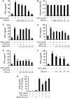
Norepinephrine (NE) inhibits lipopolysaccharide (LPS)-induced tumour necrosis factor-α production via activating α1 adrenoceptor in neonatal rat cardiomyocytes. (A, B and F) Cardiomyocytes were treated with NE, phenylephrine (PE) or vehicle for 10 min. and then with LPS or normal saline for 6 hrs. (C–E and G) After pre-treatment with prazosin (PRAZ), atenolol (ATEN) or ICI-118,551 (ICI) for 30 min., cardiomyocytes were stimulated with NE for 10 min. and with LPS for another 6 hrs (C–E) or 1.5 hrs (G). Data are mean ± SEM from four independent experiments. **P < 0.01 versus control, #P < 0.05, ##P < 0.01 versus LPS group, ΔP < 0.05 versus LPS+NE group.
Fig 2
Fig 2
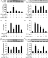
Effects of norepinephrine (NE) and prazosin (PRAZ) on lipopolysaccharide (LPS)-induced JNK1/2, p38 and extracellular signal-regulated kinase 1/2 (ERK1/2) phosphorylation and c-Fos expression in neonatal rat cardiomyocytes. After pre-treatment with PRAZ or vehicle for 30 min., cardiomyocytes were incubated with NE or vehicle for 10 min. and then with LPS or normal saline for another 30 min. Representative blots and quantification of JNK1/2 (A), p38 (B) and ERK1/2 (C) phosphorylation and c-Fos (D) expression are shown. Data are expressed as mean ± SEM, n = 5. *P < 0.05, **P < 0.01 versus control group, #P < 0.05, ##P < 0.01 versus LPS group, ΔP < 0.05, ΔΔP < 0.01 versus LPS+NE group.
Fig 3
Fig 3
Effects of norepinephrine (NE) and prazosin (PRAZ) on lipopolysaccharide (LPS)-induced NF-κB activation in neonatal rat cardiomyocytes. Cardiomyocytes were treated as described in Figure 2. (A) NF-κB p65 nuclear translocation was analysed by laser confocal microscopy. Scale bar = 20 μm. (B and C) The cytosolic and nuclear NF-κB p65 levels were assessed by western blot; data are expressed as mean ± SEM, n = 5. *P < 0.05, **P < 0.01 versus control, #P < 0.05, ##P < 0.01 versus LPS group, ΔP < 0.05, ΔΔP < 0.01 versus LPS+NE group.
Fig 4
Fig 4
Norepinephrine (NE) enhances c-Fos expression, inhibits p38 mitogen-activated protein kinase and in turn partly decreased tumour necrosis factor α (TNF-α) production, but not NF-κB activation, via activating extracellular signal-regulated kinase 1/2 (ERK1/2) signal pathway in lipopolysaccharide (LPS)-challenged cardiomyocytes. After pre-treatment with ERK1/2 inhibitor (U0126), p38 inhibitor (SB 202190) or vehicle for 30 min., cardiomyocytes were stimulated with NE or vehicle for 10 min. and then exposed to LPS or normal saline for additional 30 min. (A, B, E and F) or 6 hrs (C and D). Expression of c-Fos (A), p38 phosphorylation (B), cytosolic (E) and nuclear (F) NF-κB p65 levels were determined by western blot. TNF-α level in the supernatant was detected by ELISA (C and D). Data are mean ± SEM, n = 5–6. **P < 0.01 versus control, #P < 0.05, ##P < 0.01 versus LPS group, ΔP < 0.05, ΔΔP < 0.01 versus LPS+NE group.
Fig 5
Fig 5
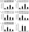
Effects of α1-AR agonists, phenylephrine (PE), on lipopolysaccharide (LPS)-induced myocardial extracellular signal-regulated kinase 1/2 (ERK1/2), p38 and IκBα phosphorylation, c-Fos expression as well as myocardial and plasma tumour necrosis factor α (TNF-α) production in mice. BALB/c mice were challenged with LPS (20 mg/kg), and PE (20 μg/kg) was injected subcutaneously 30 min. before and 2 hrs after LPS administration respectively. At 2.5 hrs after LPS administration, myocardial ERK1/2 (A), p38 (C) and IκB (D) phosphorylation, c-Fos expression (B), myocardial (E) and plasma (F) TNF-α levels were examined by western blot or ELISA. Data are mean ± SEM, n = 8. *P < 0.05, **P < 0.01 versus control, #P < 0.05, ##P < 0.01 versus LPS group.
Fig 6
Fig 6
Effect of phenylephrine (PE) on cardiac function in endotoxaemic mice. Mice were challenged with LPS (20 mg/kg), and PE (5, 10 or 20 μg/kg) was injected subcutaneously 30 min. before and 2 hrs after LPS administration respectively. (A) The representative M-mode echocardiograms at 12 hrs after LPS administration. (B) LV ejection fraction (EF), (C) fractional shortening (FS), (D) stroke volume (SV) and (E) cardiac output (CO) are presented. Data are mean ± SEM, n = 7–10. **P < 0.01 versus control, #P < 0.05, ##P < 0.01 versus LPS group.

Similar articles

Cited by

References

    1. Fernandes CJ, Jr, de Assuncao MS. Myocardial dysfunction in sepsis: a large, unsolved puzzle. Crit Care Res Pract. 2012;2012:896430. Doi: 10.1155/2012/896430. - DOI - PMC - PubMed
    1. Zhang T, Feng Q. Nitric oxide and calcium signaling regulate myocardial tumor necrosis factor-α expression and cardiac function in sepsis. Can J Physiol Pharmacol. 2010;88:92–104. - PubMed
    1. Natanson C, Eichenholz PW, Danner RL, et al. Endotoxin and tumor necrosis factor challenges in dogs simulate the cardiovascular profile of human septic shock. J Exp Med. 1989;169:823–32. - PMC - PubMed
    1. Cain BS, Meldrum DR, Dinarello CA, et al. Tumor necrosis factor-alpha and interleukin-1beta synergistically depress human myocardial function. Crit Care Med. 1999;27:1309–18. - PubMed
    1. Vincent JL, Bakker J, Marécaux G, et al. Administration of anti-TNF antibody improves left ventricular function in septic shock patients. Results of a pilot study. Chest. 1992;101:810–5. - PubMed

Publication types

MeSH terms