. 2013 Dec 5:10:147.
doi: 10.1186/1742-4690-10-147.

Allosteric induction of the CD4-bound conformation of HIV-1 Gp120

Affiliations

Allosteric induction of the CD4-bound conformation of HIV-1 Gp120

Anna Roitburd-Berman et al. Retrovirology. .

Abstract

Background: HIV-1 infection of target cells is mediated via the binding of the viral envelope protein, gp120, to the cell surface receptor CD4. This interaction leads to conformational rearrangements in gp120 forming or revealing CD4 induced (CD4i) epitopes which are critical for the subsequent recognition of the co-receptor required for viral entry. The CD4-bound state of gp120 has been considered a potential immunogen for HIV-1 vaccine development. Here we report on an alternative means to induce gp120 into the CD4i conformation.

Results: Combinatorial phage display peptide libraries were screened against HIV-1 gp120 and short (14aa) peptides were selected that bind the viral envelope and allosterically induce the CD4i conformation. The lead peptide was subsequently systematically optimized for higher affinity as well as more efficient inductive activity. The peptide:gp120 complex was scrutinized with a panel of neutralizing anti-gp120 monoclonal antibodies and CD4 itself, illustrating that peptide binding does not interfere with or obscure the CD4 binding site.

Conclusions: Two surfaces of gp120 are considered targets for the development of cross neutralizing antibodies against HIV-1; the CD4 binding site and CD4i epitopes. By implementing novel peptides that allosterically induce the CD4i epitopes we have generated a viral envelope that presents both of these surfaces simultaneously.


Figures
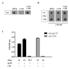
Figure 1
Figure 1
The m1-phage binds gp120 and locks it into a CD4i conformation without interfering with CD4 binding. A. Dot blot illustrating the stringent dependence of mAb CG10 on CD4. CDC451 gp120 was spotted onto nitrocellulose and detected with HIVIg, mAb CG10 or mAb CG10 in the presence of CD4. CG10 is strictly dependent on the presence of CD4 for gp120 binding. B. Equal amounts of phages displaying the m1-peptide, phages with no insert (fth-1) or CD4 were spotted to nitrocellulose filters. Filters were incubated with gp120 CDC451 or in blocking solution without gp120 (no signals developed in the absence of gp120, not shown) and bound gp120 was detected using HIVIg or the CD4i CG10 mAb. C. Equal amounts of phages displaying the m1-peptide or phages with no insert (fth-1) were captured on ELISA wells using an anti-M13 mAb. Wells were incubated with pre-formed gp120 CDC451:CD4 complex (stoichiometric molar ratio of 1:1), gp120 or CD4 alone, as indicated. Bound gp120 was detected using a biotinylated anti-gp120 9G3 mAb; bound CD4 was detected by a biotinylated anti-CD4 mAb CG9. The experiment was carried out in triplicate.
Figure 2
Figure 2
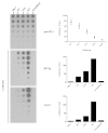
Semi-quantitative dot blot analyses of m2-phage binding to gp120. Equal amounts of 5 different phages were applied to nitrocellulose filters (two fold serial dilutions) and reacted with rabbit anti-M13 polyclonal sera, or gp120 CDC451 followed by HIVIg or CG10 mAb as indicated. As is illustrated in the top filter, the amount of phages at each dilution for each of the five different phages is similar and the ECL signal drops as the phages are diluted (see densitometric quantification in the histogram on the right and Methods). The binding of gp120 and detection with CG10 is enhanced for phage 2A6 (obtained in standard biopanning) and markedly improved for m2-phage (obtained in stringent biopanning) compared to phage m1, as is detected in the filters and their subsequent densitometric scans (histograms on the right) which were performed for both HIVIg and CG10 at dilution 1:4. Peptide sequences: m1 – C-DRRDLPQWAKRE-C; 2A6 – C-DRRDLPQWAIRE-C; m2- C-DRRDLPDWAIRA-C; scrambled – C-DLWRIRADRAPD-C; fth-1 – no insert.
Figure 3
Figure 3
The m2-phage binds monomeric and trimeric envelope proteins of different HIV-1 isolates. Equal amounts of phages and CD4 were applied to nitrocellulose filters and incubated with gp120 CDC451, gp120 BaL, trimeric R2 gp140 or without any envelope as indicated (no signals developed in the absence of envelope, not shown). Captured envelope proteins were detected using HIVIg or the CD4i mAb CG10.
Figure 4
Figure 4
The CD4 binding site is not compromised by m2-phage binding. The m2 and fth-1 phages were applied to nitrocellulose filters along with CD4. The filters were incubated with gp120 BaL or without gp120 (no signals developed in the absence of gp120, not shown) and probed with three stringent CD4i mAbs and three CD4bs mAbs as indicated. Binding is also illustrated for mAb 2G12 which recognizes a mannose-rich epitope on gp120. Note that m2-phage induces binding for all three of the CD4i mAbs and does not interfere with the binding of the CD4bs mAbs. The binding of the CD4bs mAbs is sensitive to the capture of gp120 by CD4 as expected.
Figure 5
Figure 5
The binding sites for CD4 and m2-phage are different. CD4 was applied to ELISA wells and used to capture gp120 BaL. Subsequently m2-phage was added and detection of phage and gp120 was achieved with anti-M13 mAb, HIVIg or b12 mAb as indicated. Note that whereas CD4 capture inhibits b12 binding as expected, occupation of the CD4bs does not interfere with m2 binding. The experiment was carried out in duplicate.
Figure 6
Figure 6
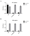
Comparative capture of gp120 with two defining mAbs. gp120 BaL was captured on ELISA wells using immobilized b12 mAb (A) compared with immobilized 1B6 mAb (B) and incubated with different phages as indicated. Bound phages were detected with rabbit anti-M13 polyclonal sera while detection of gp120 was accomplished using the LG4 mAb. Note that both b12 and 1B6 are efficient in capturing gp120 BaL yet distinct regarding overlap with the m2 binding epitope. The b12 epitope overlaps the CD4bs (see for example Figure 5) and does not interfere with m2 binding. The 1B6 epitope is different and distinct, competes for m2 binding and does not interfere with binding of either CD4 or b12 (see Figure 7). The experiment was carried out in duplicate.
Figure 7
Figure 7
Mutagenesis of the CD4 binding site does not alter the m2 binding site. Wild type gp120 BaL or the gp120 BaL D368R mutant were captured onto ELISA wells using mAb 1B6 or HIVIg as indicated. The captured gp120 was then detected with various anti-gp120 mAbs (as indicated). CD4 binding to captured gp120 was detected with biotinylated anti-CD4 mAb CG9. Bound phages were detected with an anti-M13 mAb. Note the 1B6 mAb which competes with m2 binding (Figure 6) does not interfere with anti-gp120 mAbs or CD4. The experiment was carried out in duplicate.
Figure 8
Figure 8
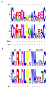
Analysis of gp120 CDC451-binding peptides affinity selected from the biased random mutagenesis library and analysis of the “X6 NNK” m2-based phage display library. A.Analysis of the biased random mutagenesis library. 24 randomly selected phages from the biased random mutagenesis m1-based library and 23 affinity selected phages obtained after screening of the biased random mutagenesis m1-based library against gp120 CDC451 were sequenced. The sequences were used as input for the MEME Suite motif analysis software [60,61] and two logos were produced (random peptides top and affinity-selected peptides bottom). The sequences of the m1, 2A6 (standard screening) and m2 (stringent biopaning) peptides are provided for comparison. B.Analysis of the “X6 NNK” m2-based library. Logos were prepared from the sequences of 20 randomly sampled phages of the “X6 NNK” m2-based library (upper logo) and those of 41 affinity selected phages obtained by screening the library with envelope as described in the text (lower logo). The sequence of m3-peptide is provided for comparison.
Figure 9
Figure 9
SPR analyses of gp120 CDC451 binding to peptides. Biotinylated phages displaying peptides m1, m2 and m3 were immobilized on CM5 sensor chips coated with streptavidin and reacted with either gp120 (red), mAb CG10 (blue) or with the mixture gp120 + CG10 (black). Y-axes (Signal) were adjusted according to baseline. As can be seen, the binding to m2-phage is markedly improved as compared to m1. The m3 peptide binds marginally better than m2. A scrambled peptide which was also tested in this setting did not show any binding to gp120 or CG10 (not shown). The two right-hand panels depict binding kinetics of two fold serial dilutions of gp120 (3.9-250nM) to the immobilized m2 displaying phage. The experimental data (colored curves) were fitted (black curves) using TraceDrawer 1.5 software (Ridgeview Instruments AB Uppsala, Sweden). As can be seen in A the OneToOne model does not fit the data very well (Chi2 4.25). Using the OneToOne TwoState model the fit is markedly improved (Chi2 0.32) supporting the conclusion that binding of m2-phage to gp120 is associated with conformational rearrangements.

Similar articles

Cited by

References

    1. Dalgleish AG, Beverley PC, Clapham PR, Crawford DH, Greaves MF, Weiss RA. The CD4 (T4) antigen is an essential component of the receptor for the AIDS retrovirus. Nature. 1984;312:763–767. doi: 10.1038/312763a0. - DOI - PubMed
    1. Klatzmann D, Champagne E, Chamaret S, Gruest J, Guetard D, Hercend T, Gluckman JC, Montagnier L. T-lymphocyte T4 molecule behaves as the receptor for human retrovirus LAV. Nature. 1984;312:767–768. doi: 10.1038/312767a0. - DOI - PubMed
    1. DeVico AL. CD4-induced epitopes in the HIV envelope glycoprotein, gp120. Curr HIV Res. 2007;5:561–571. doi: 10.2174/157016207782418560. - DOI - PubMed
    1. Kong L, Huang CC, Coales SJ, Molnar KS, Skinner J, Hamuro Y, Kwong PD. Local conformational stability of HIV-1 gp120 in unliganded and CD4-bound states as defined by amide hydrogen/deuterium exchange. J Virol. 2010;84:10311–10321. doi: 10.1128/JVI.00688-10. - DOI - PMC - PubMed
    1. Mao Y, Wang L, Gu C, Herschhorn A, Xiang SH, Haim H, Yang X, Sodroski J. Subunit organization of the membrane-bound HIV-1 envelope glycoprotein trimer. Nat Struct Mol Biol. 2012;19:893–899. doi: 10.1038/nsmb.2351. - DOI - PMC - PubMed

Publication types

Substances

LinkOut - more resources