Review
. 2013 Dec;19(6 Muscle Disease):1509-34.
doi: 10.1212/01.CON.0000440658.03557.f1.

Congenital muscular dystrophies and congenital myopathies

Review

Congenital muscular dystrophies and congenital myopathies

Susan T Iannaccone et al. Continuum (Minneap Minn). 2013 Dec.

Abstract

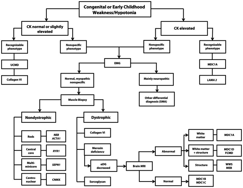
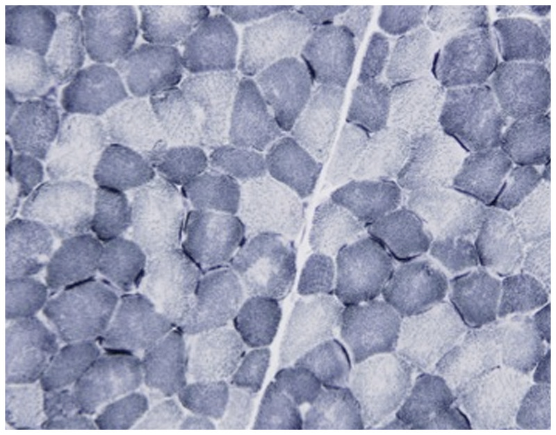
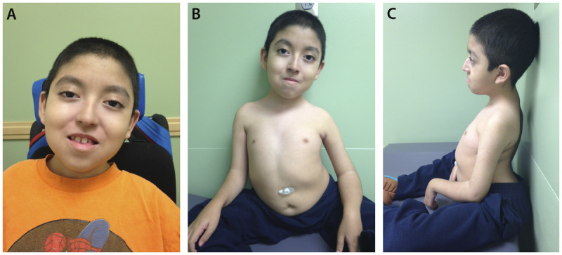
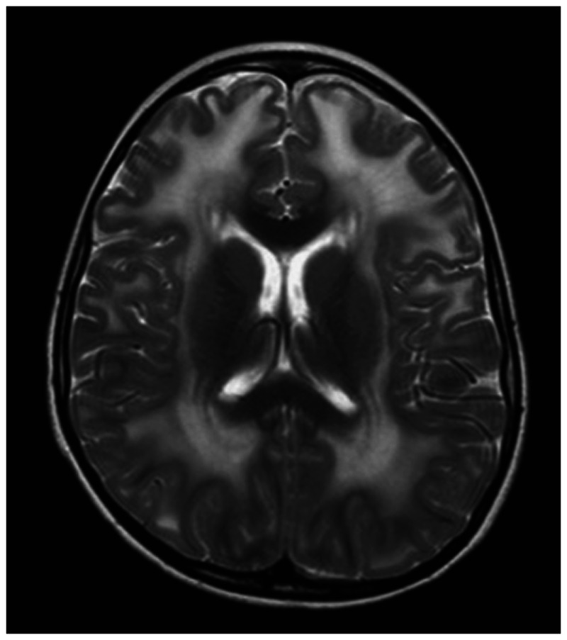
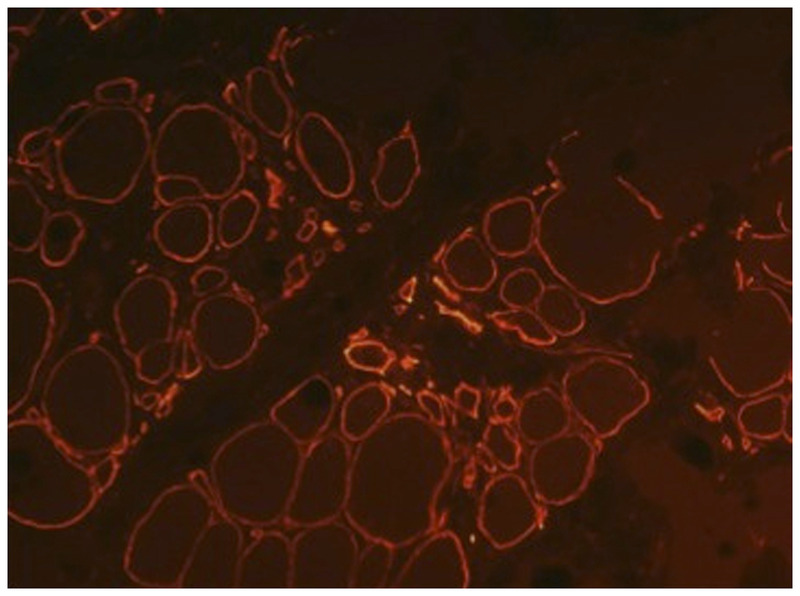
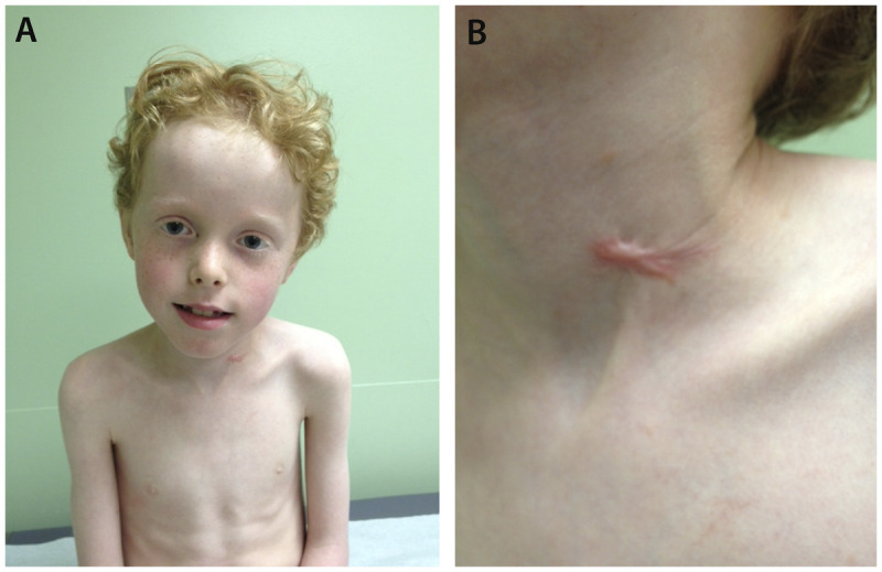
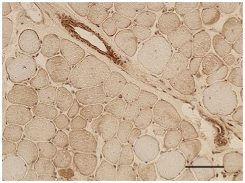
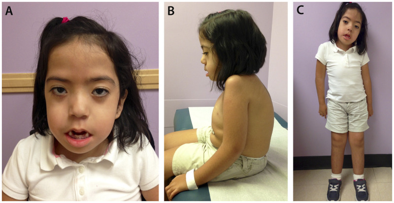
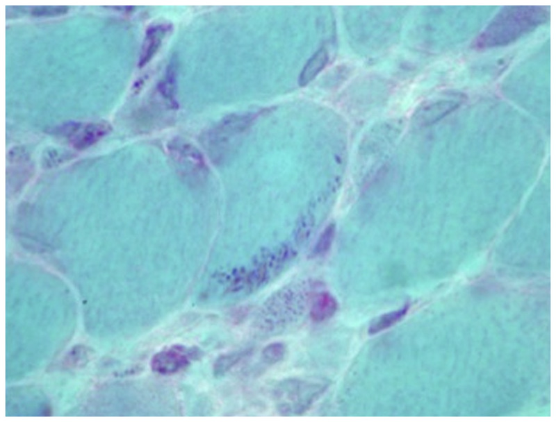
Purpose of review: The purpose of this review is to provide information regarding the diagnosis and natural history of some very rare disorders: congenital muscular dystrophies and congenital myopathies. Patients with these conditions share characteristics such as early onset of weakness and severe hypotonia. Other organs such as the brain, eyes, and skin may be involved. Diagnosis depends largely on recognition of phenotype, muscle biopsy, and mutation analysis.

Recent findings: More than 30 genes have been associated with these diseases, most of which have only been recognized in the past decade. Increasing availability of DNA analysis has been important in decreasing delay in diagnosis.

Summary: Patients with congenital muscular dystrophy or congenital myopathy are at high risk of complications including restrictive lung disease, orthopedic deformities, seizures, cardiomyopathy, and malignant hyperthermia. Life expectancy varies with the severity of complications. Having an accurate and specific diagnosis allows the neurologist to carry out anticipatory guidance and appropriate monitoring. New hope exists for experimental treatments for congenital muscular dystrophy and congenital myopathy as our understanding of pathogenesis evolves.


Figures

Figure 1-1.
Figure 1-1.
Hematoxylin and eosin–stained section of left quadriceps demonstrates hypertrophic fibers with sarcoplasmic disarray interspersed with atrophic fibers, some with sarcoplasmic basophilia consistent with regeneration. Occasional inflammatory cells are in the interstitial space (200×).
Figure 1-2.
Figure 1-2.
Algorithm for the diagnosis of congenital muscular dystrophy. CK = creatine kinase; UCMD = Ullrich congenital muscular dystrophy; EMG = electromyography; MDC1A = merosin-deficient congenital muscular dystrophy; LAMA 2 = laminin α2 gene; SMA = spinal muscular atrophy; NEB = nebulin gene; ACTA1 = gene encoding skeletal muscle actin; RYR1 = ryanodine receptor; MDC1D = congenital muscular dystrophy type 1D; FCMD = Fukuyama congenital muscular dystrophy; SEPN1 = selenoprotein 1; αDG = α-dystroglycanopathy; MRI = magnetic resonance imaging; WWS = Walker-Warburg syndrome; MEB = muscle-eye-brain disease; CNMX = X-linked centronuclear myopathy; MDC1B = congenital muscular dystrophy type 1B; MDC1C = congenital muscular dystrophy type 1C.
Figure 1-10.
Figure 1-10.
Electron microscopy showing minicores affecting focal areas of the fiber. A myofibrillary disruption and paucity of mitochondria is shown, as well as degeneration of the sarcomere and transverse tubules.
Figure 1-11.
Figure 1-11.
Central core myopathy. NADH–tetrazolium reductase–stained section of left quadriceps showing predominant type 1 fibers with cores affecting the majority of fibers. Cores extend along the fiber length on longitudinal sections and are typically well demarcated. Courtesy of Emily Herndon, MD.
Figure 1-12.
Figure 1-12.
Multiminicore disease. NADH–tetrazolium reductase–stained section of left quadriceps demonstrating multiple areas in both fiber types of varying size and number devoid of oxidative enzyme stain compatible with multiminicore structures. Minicores are usually perpendicular to the longitudinal axis. Courtesy of Emily Herndon, MD.
Figure 1-3.
Figure 1-3.
An 8-year-old boy with merosin-deficient congenital muscular dystrophy confirmed by DNA mutation analysis, which revealed a mutation of the LAMA 2 gene. A, Evidence of bilateral ptosis and facial weakness; B, severe dextroscoliosis and the presence of a gastrostomy tube; C, hyperlordosis and contractures at the elbow and wrist.
Figure 1-4.
Figure 1-4.
Brain MRI with diffuse prolonged T2 signal seen in the deep and subcortical white matter through both cerebral hemispheres in a 10-year-old boy with merosin-deficient congenital muscular dystrophy.
Figure 1-5.
Figure 1-5.
Merosin-deficient congenital muscular dystrophy (MDC1A). Immunohistochemical stain with an antibody to laminin α2 showing partial expression of merosin (200×).
Figure 1-6.
Figure 1-6.
A 9-year-old boy with Ullrich congenital muscular dystrophy. A, Patient shows muscle wasting and bilateral elbow contractures and, B, abnormal keloid formation.
Figure 1-7.
Figure 1-7.
Dystroglycanopathy. Immunohistochemical stain with an antibody to α-dystroglycan reveals attenuated sarcolemmal reactivity in most fibers (200×). Courtesy of Emily Herndon, MD.
Figure 1-8.
Figure 1-8.
A 7-year-old girl with nemaline myopathy diagnosed by muscle biopsy. A, Myopathic face with severe facial weakness and micrognathia; B, chest wall deformity, kyphosis, and gastrostomy tube; C, dextroscoliosis and muscle atrophy more notable in the upper extremities.
Figure 1-9.
Figure 1-9.
Nemaline myopathy. Gomori one-step trichrome-stained section of left quadriceps showing multiple clusters of red staining rods at the periphery of most fibers, which is compatible with nemaline rods (400×). Courtesy of Emily Herndon, MD.

References

    1. Tome FM,, Evangelista T,, Leclerc A, et al. Congenital muscular dystrophy with merosin deficiency. CR Acad Sci III 1994; 317 (4): 351–357. - PubMed
    1. Clement EM,, Feng L,, Mein R, et al. Relative frequency of congenital muscular dystrophy subtypes: analysis of the UK diagnostic service 2001-2008. Neuromuscul Disord 2012; 22 (6): 522–527. - PubMed
    1. Peat RA,, Smith JM,, Compton AG, et al. Diagnosis and etiology of congenital muscular dystrophy. Neurology 2008; 71 (5): 312–321. - PubMed
    1. Okada M,, Kawahara G,, Noguchi S, et al. Primary collagen VI deficiency is the second most common congenital muscular dystrophy in Japan. Neurology 2007; 69 (10): 1035–1042. - PubMed
    1. Geranmayeh F,, Clement E,, Feng LH, et al. Genotype-phenotype correlation in a large population of muscular dystrophy patients with LAMA2 mutations. Neuromuscul Disord 2010; 20 (4): 241–250. - PubMed

Publication types

MeSH terms