. 2014 Apr;13(2):292-302.
doi: 10.1111/acel.12173. Epub 2014 Jan 24.

Embryonic expression of the common progeroid lamin A splice mutation arrests postnatal skin development

Affiliations

Embryonic expression of the common progeroid lamin A splice mutation arrests postnatal skin development

Tomás McKenna et al. Aging Cell. 2014 Apr.

Abstract

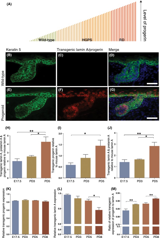
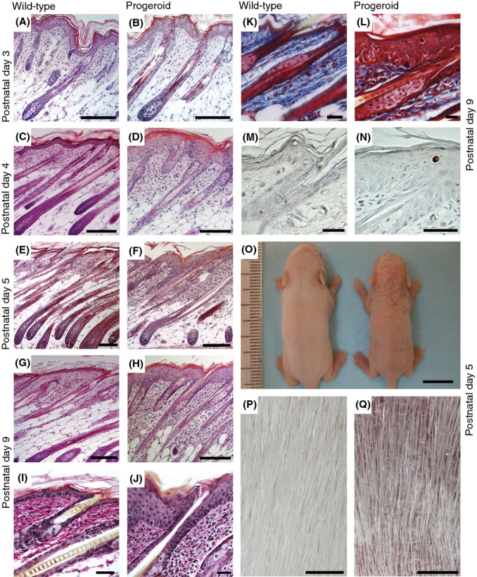
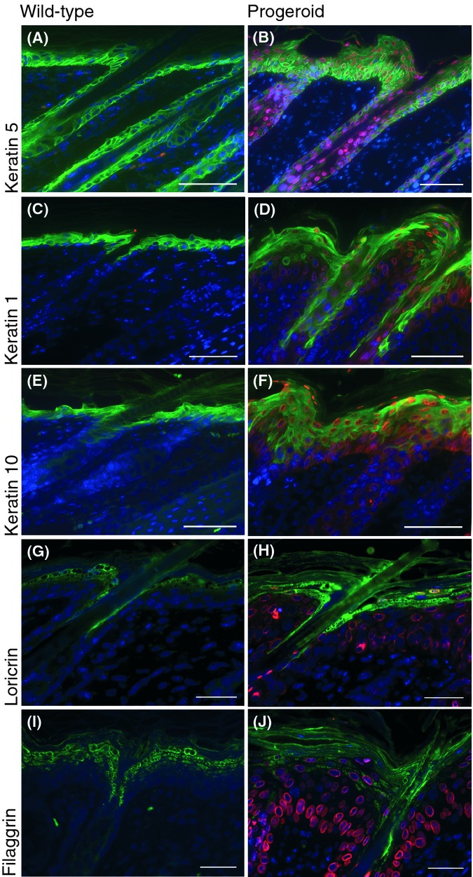
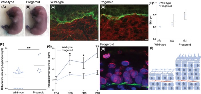
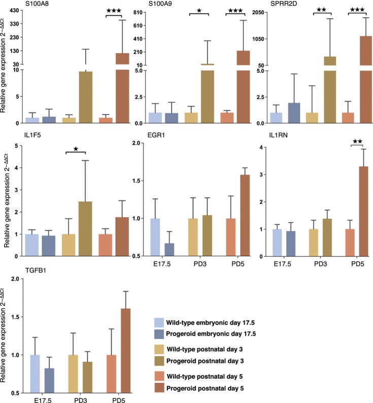
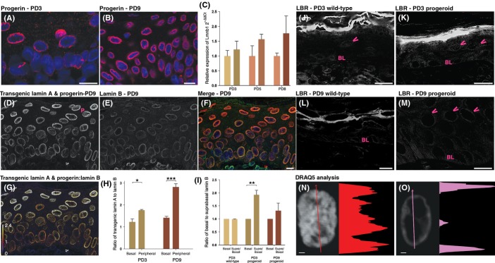
Hutchinson-Gilford progeria syndrome (HGPS) and restrictive dermopathy (RD) are two laminopathies caused by mutations leading to cellular accumulation of prelamin A or one of its truncated forms, progerin. One proposed mechanism for the more severe symptoms in patients with RD compared with HGPS is that higher levels of farnesylated lamin A are produced in RD. Here, we show evidence in support of that hypothesis. Overexpression of the most common progeroid lamin A mutation (LMNA c.1824C>T, p.G608G) during skin development results in a severe phenotype, characterized by dry scaly skin. At postnatal day 5 (PD5), progeroid animals showed a hyperplastic epidermis, disorganized sebaceous glands and an acute inflammatory dermal response, also involving the hypodermal fat layer. PD5 animals also showed an upregulation of multiple inflammatory response genes and an activated NF-kB target pathway. Careful analysis of the interfollicular epidermis showed aberrant expression of the lamin B receptor (LBR) in the suprabasal layer. Prolonged expression of LBR, in 14.06% of the cells, likely contributes to the observed arrest of skin development, clearly evident at PD4 when the skin had developed into single-layer epithelium in the wild-type animals while progeroid animals still had the multilayered appearance typical for skin at PD3. Suprabasal cells expressing LBR showed altered DNA distribution, suggesting the induction of gene expression changes. Despite the formation of a functional epidermal barrier and proven functionality of the gap junctions, progeroid animals displayed a greater rate of water loss as compared with wild-type littermates and died within the first two postnatal weeks.

Keywords: HGPS; aging; lamin B; lamin B receptor; progerin; restrictive dermopathy.


Figures

Figure 1
Figure 1
Overexpression of the common progeroid mutation, LMNA c.1824C>T; p. G608G, during skin development. One proposed mechanism for the more severe symptoms in patients with RD compared with HGPS is that higher levels of the progerin protein are produced in RD, and as progerin levels increase, the disease severity also increases (A). Transgenic lamin A and progerin expression were detected in the hair follicles and in the interfollicular epidermis by immunofluorescent staining of skin sections using an antibody against keratin 5 and human lamin A/C in wild-type (B–D) and progeroid (E–G) E17.5 embryos. The transgenic expression of lamin A and progerin follows the keratin 5 expression in progeroid embryos (E–G). No staining with the human lamin A/C antibody was detected in wild-types (C). The sizes of the proteins expressed from the transgene were further confirmed using Western blot. The relative transgenic overexpression was quantified using densitometry from Western blot filters hybridized with an antibody that recognized lamin A of both human and mouse origin (H–J). Trangenic expression was further evaluated using qPCR with primers specific for progerin (K), and human lamin A (L), normalized to β-actin. The ratio of normalized transgenic progerin to human lamin (M) suggests an increase in progerin but is actually reflecting the drop in lamin A expression in (L). A break in the Y-axis designates a change in the scale. N ≥ 3 for both wild-types and progeroid animals per age group. Error bars indicate SEM, *P < 0.05, **P < 0.01). Scale bars indicate 200 μm.
Figure 2
Figure 2
Embryonic overexpression of the LMNA c.1824C>T; p. G608G mutation results in a severe postnatal skin phenotype. Histological examination of hematoxylin and eosin stained skin sections (A–H) from a PD3 wild-type (A) and progeroid animals (B) showed no visible abnormalities. Skin sections from 4-day-old wild-type animal showed the first signs of skin maturation (C), while 4-day-old progeroid skin sections showed no such signs of maturation (D). In 5-day-old animals, a skin pathology with hyperplastic epidermis, disorganized sebaceous glands, and an acute inflammatory dermal response, also involving the hypodermal fat layer, was apparent in animals expressing the LMNA c.1824 C>T mutation (F), not seen in wild-type animals (E). This skin pathology was even more pronounced in the 9-day-old progeroid skin, (H) compared with wild-type animals (G). Van Gieson staining of skin from wild-type (I) and progeroid animals (J) showed no significant difference in elastin fibers. Staining the skin using Masson’s trichrome (K and L) revealed dense collagen fibers in the progeroid section (L) not evident in the wild-type (K). Immunohistochemistry with cleaved caspase 3 (M and N), which indicates cells undergoing apoptosis, did not show a significant increase in the number of apoptotic cells in skin from progeroid (N) compared with wild-type (M) animals. A photograph of a 5-day-old progerin expressing animal and a wild-type littermate revealed a diminished size, as well as hair thinning and dry scaly skin (O), which were highlighted in (P) and (Q). Scale bars for (A–H) indicate 100 μm, for (O) 1 cm, for (P and Q) 1 mm, for (I–N) 20 μm.
Figure 3
Figure 3
Markers for epidermal differentiation were expressed in progeroid skin. Epidermal differentiation markers (in green) in dorsal skin sections from 5-day-old wild-type and progeroid animals. Immunofluorescent staining for keratin 5 (A and B) showed, as expected, positive cells in the basal cell layer of the interfollicular epidermis, the outer root sheath, and the peripheral cells of the sebaceous gland; however, in progeroid sections, keratin 5 was also aberrantly expressed in suprabasal layers of the epidermis. Keratin 1 (C and D) and keratin 10 (E and F) were found in the suprabasal layers of the epidermis, mostly in the spinous layer. Loricrin (G and H) and filaggrin (I and J) antibodies localized positive cells in the granular layer of the epidermis. Transgenic human lamin A and progerin expression in progeroid sections was detected with the human specific lamin A/C antibody, shown in red. DAPI is shown in blue. Scale bars indicate 200 μm.
Figure 4
Figure 4
No evidence of delayed epidermal barrier formation, although transepidermal water loss is increased in progeroid animals. Photographs of embryos (E17.5) stained with toluidine blue show no difference between wild-type (A) and progerin expressing (B) embryos. Immunofluorescent stainings of dorsal skin sections from postnatal day 2 animals were made to examine the function of the tight junction layer (C–D). The tight junction layer protected against egress of the biotin in the progeroid as in the wild-type (C and D). Skin pH was measured from the day of birth until PD2 to ascertain whether skin acidification was affected in progeroid animals; however, skin acidification was unaffected (E). The outward barrier function was tested by a dehydration assay. Newborn pups were separated from their mother to prevent fluid intake and their dehydration rate was calculated by measuring the weight loss as a function of time and initial body weight. Progeroid animals showed an increased dehydration rate compared with wild-type littermates (F). Increased rate of transepidermal water loss was also seen in progeroid animals compared with their wild-type littermates (G). The greater exposure area of nucleated cells outside of the tight junction layer, due to the epidermal hyperplasia, might account for the greater rate of dehydration seen in progeroid compared with wild-type animals (H and I). N ≥ 3 for both wild-types and progeroid animals (A–H). Error bars indicate SEM. (*P < 0.05, **P < 0.01–0.001).
Figure 5
Figure 5
Upregulation of skin inflammatory factors and target of the NF-kB pathway in progeroid animals. In embryonic skin (E17.5), no inflammation markers were upregulated but starting in 3-day-old animals (PD3) an inflammation response had begun, preceding the pathologic changes, which were not evident until postnatal day 4. The inflammation response continued and increased in day 5 (PD5) animals. Normalized expression levels of NF-kB target genes (Egr1 and Il1rn) and the Tgf-b1 gene at different stages during skin development. Values represent mean and SD (*P < 0.05, **P < 0.01–0.001, ***P < 0.001).
Figure 6
Figure 6
The ratio of lamin B to transgenic lamin A and progerin decreases in the peripheral cells of the epidermis. Immunofluorescent sections of PD3 and PD9 progeroid skin using an antibody against progerin (A and B) showed lamina localization and accumulation of progerin in PD9 compared with PD3. Quantitative RT–PCR showed no significant change in lamin B1 expression between PD 3, 5, and 8 wild-type and progeroid animals (C). However, immunofluorescent sections of a progeroid PD9 animal showing transgenic lamin A and progerin (D) and lamin B (E) revealed a stark change in the ratios of transgenic lamin A and progerin to lamin B. Merged with transgenic lamin A and progerin in red, lamin B in green and DRAQ5 in blue (D and E merged in F). This relationship is highlighted in (G), which shows a ratio between these proteins, with high lamin B ratio in purple, a one-to-one ratio in red and a high transgenic lamin A and progerin ratio in white. A bar chart summarizing these data (H) shows a trend of increased transgenic lamin A and progerin compared with lamin B in the peripheral cells of the interfollicular epidermis compared with the ratio in basal cells at PD3, with a significantly increased ratio by PD9. The ratio of lamin B in suprabasal cells compared with basal cells was also significantly increased in both PD3 and PD9 progeroid animals compared with their wild-type littermates (I). Lamin B receptor (LBR) expression was detected in the interfollicular epidermis by immunofluorescent stainings of skin sections from wild-type and progeroid animals (J–M). Examples of LBR-positive cells are marked with arrowheads. The basal cell layer is indicated with BL. The peripheral cell layer is marked with a P in (D). The intensity profile of DRAQ5 staining in suprabasal cells of the interfollicular epidermis was examined in PD9 animals (N and O). The graph indicates fluorescence intensity along the arrow. Error bars indicate SD for (C), SEM for (I and H). N = 3 for both wild-type and progeroid animals (C, I, H). Scales bars for (A, B, J–M) indicate 10 μm, (F) indicate 20 μm and (N and O) indicate 1 μm. (**P < 0.01–0.001, ***P < 0.001).

References

    1. Adler AS, Sinha S, Kawahara TL, Zhang JY, Segal E, Chang HY. Motif module map reveals enforcement of aging by continual NF-kappaB activity. Genes Dev. 2007;21:3244–3257. - PMC - PubMed
    1. Capell B, Collins F. Human laminopathies: nuclei gone genetically awry. Nat. Rev. Genet. 2006;7:940–952. - PubMed
    1. Charles R-P, Guitard M, Leyvraz C, Breiden B, Haftek M, Haftek-Terreau Z, Stehle J-C, Sandhoff K, Hummler E. Postnatal requirement of the epithelial sodium channel for maintenance of epidermal barrier function. J. Biol. Chem. 2008;283:2622–2630. - PubMed
    1. De Benedetto A, Kubo A, Beck LA. Skin barrier disruption: a requirement for allergen sensitization? J. Invest. Dermatol. 2012;132:949–963. - PMC - PubMed
    1. Dean JC, Gray ES, Stewart KN, Brown T, Lloyd DJ, Smith NC, Pope FM. Restrictive dermopathy: a disorder of skin differentiation with abnormal integrin expression. Clin. Genet. 1993;44:287–291. - PubMed

Publication types

MeSH terms