. 2014 Mar;16(3):441-8.
doi: 10.1093/neuonc/not158. Epub 2013 Dec 4.

Amide proton transfer imaging of adult diffuse gliomas: correlation with histopathological grades

Affiliations

Amide proton transfer imaging of adult diffuse gliomas: correlation with histopathological grades

Osamu Togao et al. Neuro Oncol. 2014 Mar.

Abstract

Background: Amide proton transfer (APT) imaging is a novel molecular MRI technique to detect endogenous mobile proteins and peptides through chemical exchange saturation transfer. We prospectively assessed the usefulness of APT imaging in predicting the histological grade of adult diffuse gliomas.

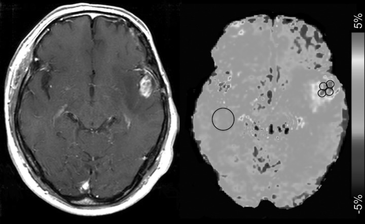
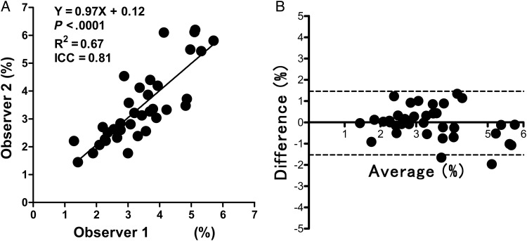
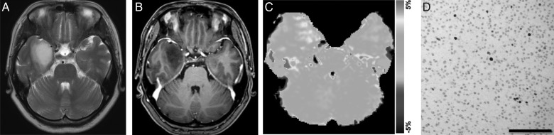
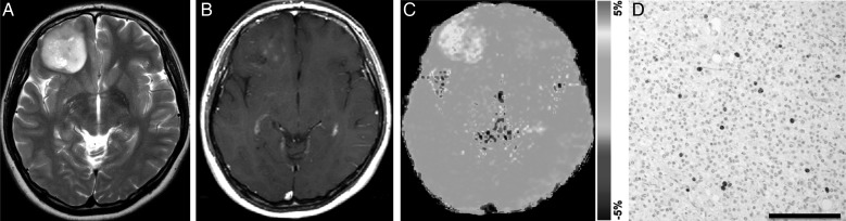
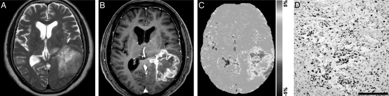
Methods: Thirty-six consecutive patients with histopathologically proven diffuse glioma (48.1 ± 14.7 y old, 16 males and 20 females) were included in the study. APT MRI was conducted on a 3T clinical scanner and was obtained with 2 s saturation at 25 saturation frequency offsets ω = -6 to +6 ppm (step 0.5 ppm). δB0 maps were acquired separately for a point-by-point δB0 correction. APT signal intensity (SI) was defined as magnetization transfer asymmetry at 3.5 ppm: magnetization transfer ratio (MTR)asym = (S[-3.5 ppm] - S[+3.5 ppm])/S0. Regions of interest were carefully placed by 2 neuroradiologists in solid parts within brain tumors. The APT SI was compared with World Health Organization grade, Ki-67 labeling index (LI), and cell density.

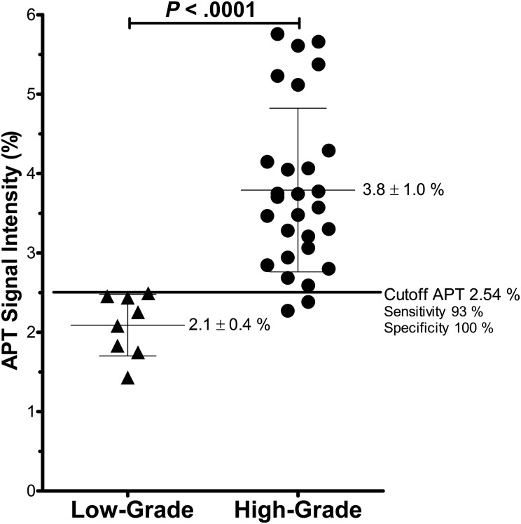
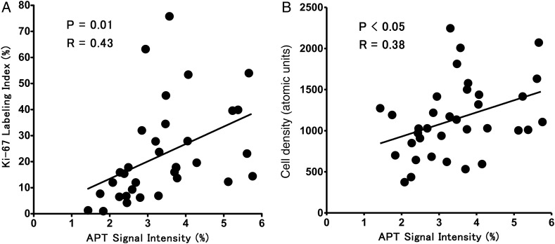
Results: The mean APT SI values were 2.1 ± 0.4% in grade II gliomas (n = 8), 3.2 ± 0.9% in grade III gliomas (n = 10), and 4.1 ± 1.0% in grade IV gliomas (n = 18). Significant differences in APT intensity were observed between grades II and III (P < .05) and grades III and IV (P < .05), as well as between grades II and IV (P < .001). There were positive correlations between APT SI and Ki-67 LI (P = .01, R = 0.43) and between APT SI and cell density (P < .05, R = 0.38). The gliomas with microscopic necrosis showed higher APT SI than those without necrosis (P < .001).

Conclusions: APT imaging can predict the histopathological grades of adult diffuse gliomas.

Keywords: amide proton transfer (APT) imaging; chemical exchange saturation transfer (CEST); glioma..


Figures

Fig. 1.
Fig. 1.
Placement of ROIs. Four circular ROIs were carefully placed in the solid component of a tumor to include the area with the highest APT signal determined by visual inspection. Cystic, necrotic, or hemorrhagic components were avoided with reference to conventional MRI. An ROI was also placed in contralateral NAWM.
Fig. 2.
Fig. 2.
Analyses of interobserver agreement. (A) The linear regression analysis shows high correlation in the APT SIs measured by the 2 observers. (B) The Bland–Altman analysis of the APT SIs measured by the 2 observers shows high concordance. Dashed lines show 95% limits of agreement.
Fig. 3.
Fig. 3.
APT SI in low-grade (grade II) and high-grade (grades III and IV) glioma. The APT SIs in the high-grade gliomas were higher than in the low-grade glioma.
Fig. 4.
Fig. 4.
Correlation between APT SI and Ki-67 LI (A) or cell density (B). Moderate positive correlations are noted between the parameters.
Fig. 5.
Fig. 5.
Astrocytoma (grade II) in a 42-year-old woman. (A) A transverse T2-weighted MRI shows a homogeneous hyperintensity area in the right temporal lobe. (B) A contrast enhanced transverse T1-weighted image shows no enhancement in the tumor. (C) The APT-weighted image shows a mild increase in SI (APT SI, 1.4%) in the tumor compared with normal brain tissue. (D) Ki-67 immunohistochemical staining shows few positive cells, indicating low proliferative activity of the tumor (Ki-67 LI, 1.3%).
Fig. 6.
Fig. 6.
Anaplastic oligodendroglioma (grade III) in a 25-year-old woman. (A) A transverse T2-weighted image shows a rather heterogeneously hyperintense area in the right frontal lobe. (B) A contrast enhanced transverse T1-weighted image shows faint enhancement in the tumor. (C) The APT-weighted image shows a mild to moderate increase in SI (APT SI, 2.8%) in the tumor compared with normal brain tissue. (D) Ki-67 immunohistochemical staining shows scattered positive cells (Ki-67 LI, 6.2%).
Fig. 7.
Fig. 7.
Glioblastoma multiforme (grade IV) in a 70-year-old woman. (A) A transverse T2-weighted image shows a heterogeneously hyperintense area in the left temporal lobe. (B) A contrast enhanced transverse T1-weighted image shows heterogeneous ringlike enhancement in the tumor. (C) The APT-weighted image shows high SI in the tumor compared with normal brain tissue (APT SI, 4.0%). (D) Ki-67 immunohistochemical staining shows a large number of positive cells, indicating high proliferative activity of the tumor (Ki-67 LI, 27.9%). High cell density is also noted.

References

    1. Louis DN, Ohgaki H, Wiestler OD, et al. The 2007 WHO classification of tumours of the central nervous system. Acta Neuropathol. 2007;114(2):97–109. - PMC - PubMed
    1. Clarke J, Butowski N, Chang S. Recent advances in therapy for glioblastoma. Arch Neurol. 2010;67(3):279–283. - PubMed
    1. Assi H, Candolfi M, Baker G, Mineharu Y, Lowenstein PR, Castro MG. Gene therapy for brain tumors: basic developments and clinical implementation. Neurosci Lett. 2012;527(2):71–77. - PMC - PubMed
    1. Dean BL, Drayer BP, Bird CR, et al. Gliomas: classification with MR imaging. Radiology. 1990;174(2):411–415. - PubMed
    1. Felix R, Schorner W, Laniado M, et al. Brain tumors: MR imaging with gadolinium-DTPA. Radiology. 1985;156(3):681–688. - PubMed

Publication types