. 2013 Dec 4;33(49):19223-37.
doi: 10.1523/JNEUROSCI.3041-13.2013.

Electrophysiological properties of rostral ventrolateral medulla presympathetic neurons modulated by the respiratory network in rats

Affiliations

Electrophysiological properties of rostral ventrolateral medulla presympathetic neurons modulated by the respiratory network in rats

Davi J A Moraes et al. J Neurosci. .

Abstract

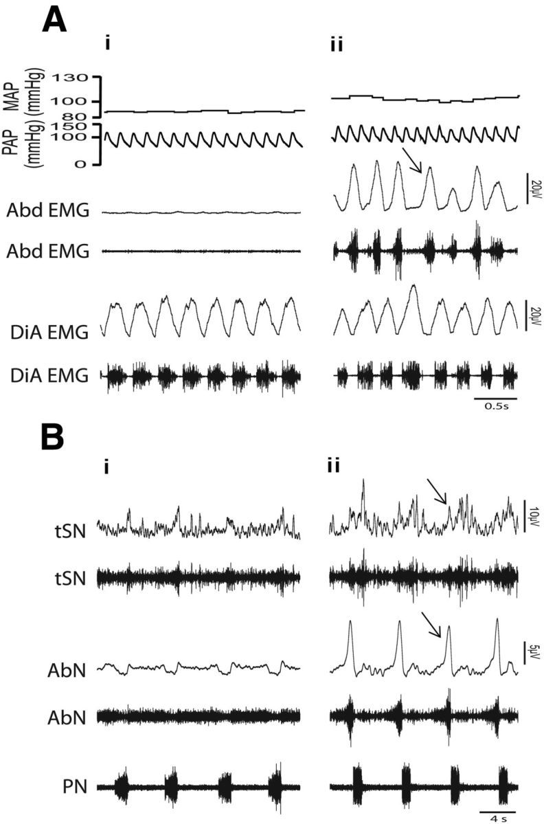
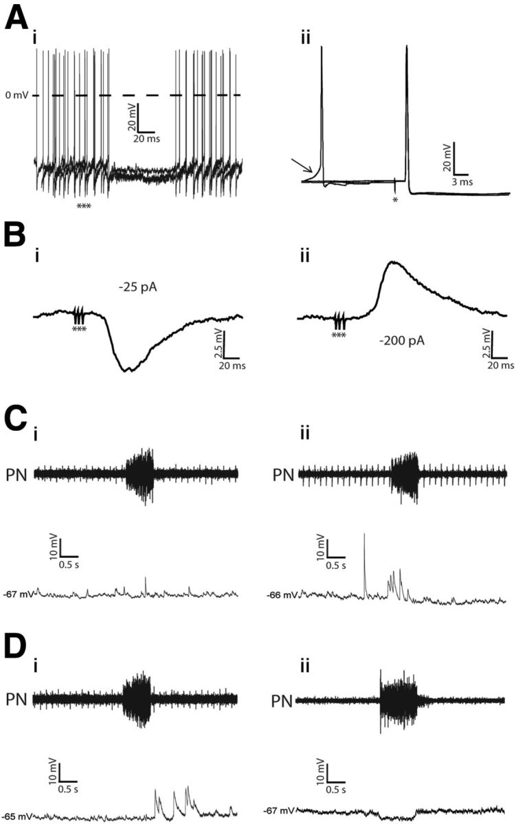
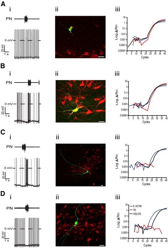
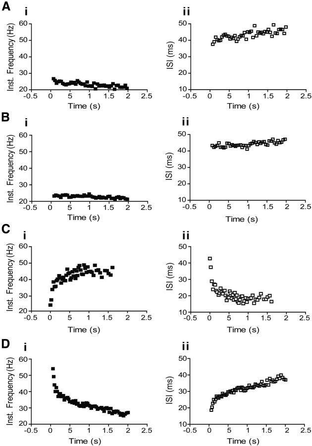
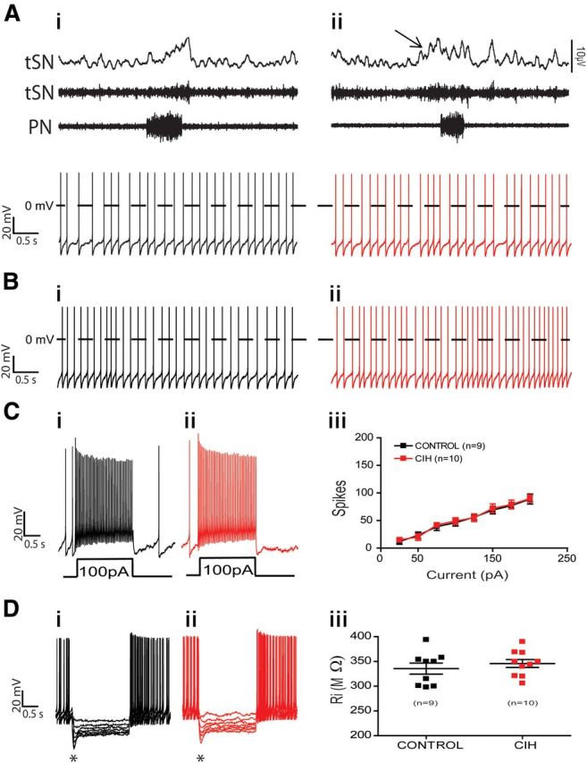
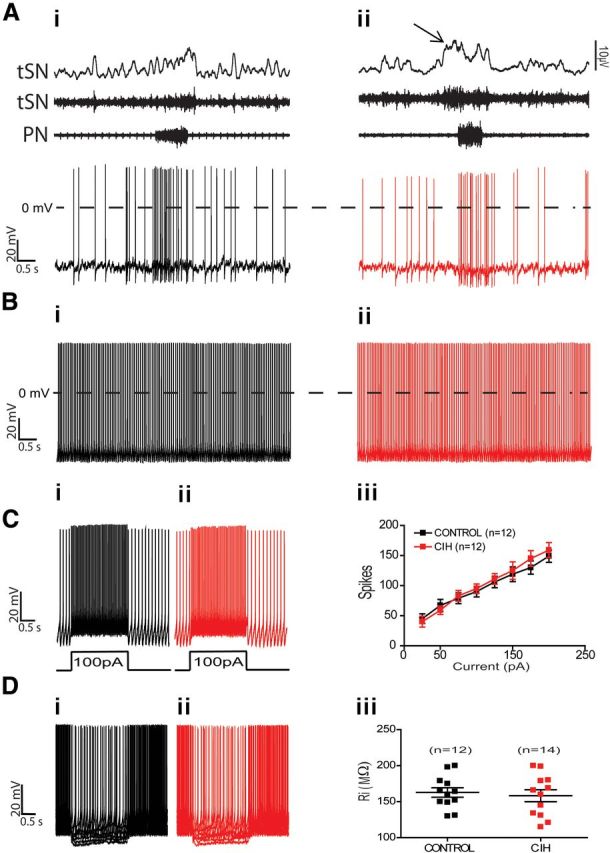
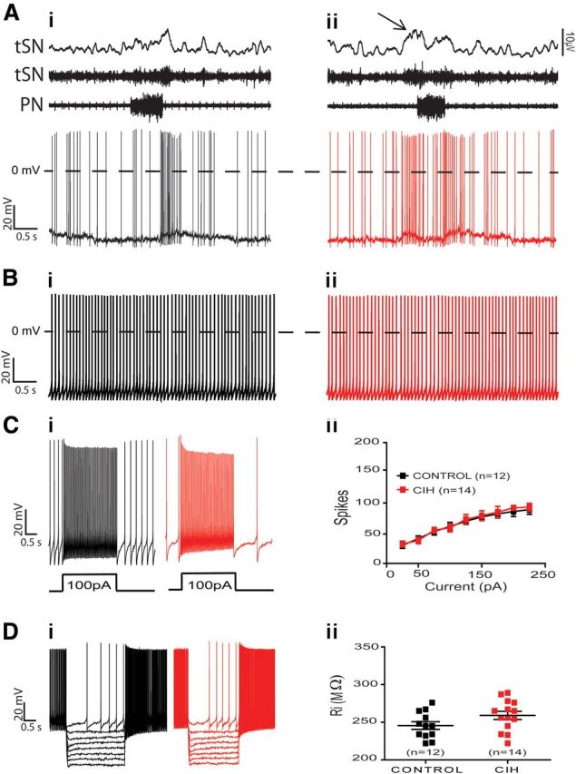
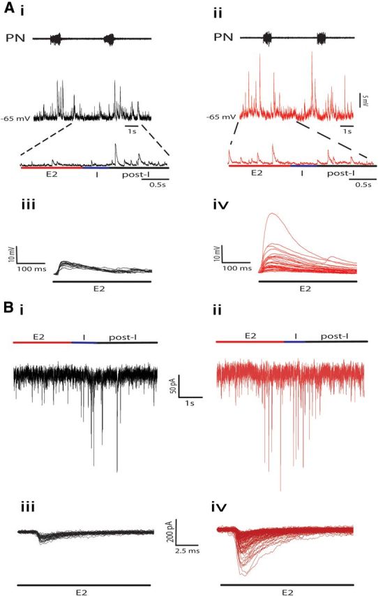
The respiratory pattern generator modulates the sympathetic outflow, the strength of which is enhanced by challenges produced by hypoxia. This coupling is due to the respiratory-modulated presympathetic neurons in the rostral ventrolateral medulla (RVLM), but the underlining electrophysiological mechanisms remain unclear. For a better understanding of the neural substrates responsible for generation of this respiratory-sympathetic coupling, we combined immunofluorescence, single cell qRT-pCR, and electrophysiological recordings of the RVLM presympathetic neurons in in situ preparations from normal rats and rats submitted to a metabolic challenge produced by chronic intermittent hypoxia (CIH). Our results show that the spinally projected cathecholaminergic C1 and non-C1 respiratory-modulated RVLM presympathetic neurons constitute a heterogeneous neuronal population regarding the intrinsic electrophysiological properties, respiratory synaptic inputs, and expression of ionic currents, albeit all neurons presented persistent sodium current-dependent intrinsic pacemaker properties after synaptic blockade. A specific subpopulation of non-C1 respiratory-modulated RVLM presympathetic neurons presented enhanced excitatory synaptic inputs from the respiratory network after CIH. This phenomenon may contribute to the increased sympathetic activity observed in CIH rats. We conclude that the different respiratory-modulated RVLM presympathetic neurons contribute to the central generation of respiratory-sympathetic coupling as part of a complex neuronal network, which in response to the challenges produced by CIH contribute to respiratory-related increase in the sympathetic activity.

Keywords: bulbospinal RVLM presympathetic neurons; chronic intermittent hypoxia; intrinsic electrophysiological properties; respiratory-sympathetic coupling.


Figures

Figure 1.
Figure 1.
Recordings of arterial pressure, respiratory muscles, and nerves in vivo and in situ preparations of control and CIH rats. Raw and integrated (∫)EMG records of inspiratory (diaphragm-DiA), expiratory muscles (abdominal-Abd) simultaneously with pulsatile arterial pressure (PAP) and mean arterial pressure (MAP) in conscious, freely moving rats from control (Ai) and CIH (Aii) rats. Note the presence of late-expiration (late-E) activity in the Abd (↑) and the increase in the MAP after CIH. Raw and integrated (∫) records of thoracic sympathetic nerve (tSN), abdominal nerve (AbN), and phrenic nerve (PN) activities of in situ preparations from the same control and CIH rats shown in Ai (Bi) and in Aii (Bii), respectively. Note the presence of late-E event (↑) in the tSN and AbN after CIH.
Figure 2.
Figure 2.
Electrophysiological identification of the nonmodulated and respiratory-modulated RVLM presympathetic neurons in in situ preparations. Ai, Hyperpolarizing potentials and inhibition of spike discharge frequency (3 superimposed sweeps, no polarizing current, membrane potential, −57 mV) evoked by stimulation of the ipsilateral ADN with bursts of 3 pulses (***). Aii, Antidromic action potentials evoked by stimulation (*) of the T8–T12 segment of the spinal cord. Three superimposed sweeps pretriggered by sEPSPs are shown. The arrow indicates spontaneous excitatory postsynaptic potential preceding spontaneous spikes. Note that spinal stimulation was followed by antidromic responses that occurred ∼6 mV more negative than the threshold at which spontaneous action potentials were induced by the ongoing synaptic activity. Bi, The ADN-evoked potentials (10 averaged sweeps) recorded intracellularly when the firing of the cell was stopped by a continuous negative current (−25 pA). Bii, Reversal of evoked inhibitory postsynaptic potential (holding current of −200 pA) evoked by stimulation of the ADN. Ci–Dii, sEPSPs, measured by hyperpolarizing the neurons, in the nonmodulated (Ci), inspiratory-modulated (Cii), postinspiratory modulated (Di), and inspiratory-inhibited (Dii) RVLM presympathetic neurons.
Figure 3.
Figure 3.
Phenotype of the nonmodulated and respiratory-modulated RVLM presympathetic neurons. Ai–Aii, Representative voltage traces of one nonmodulated RVLM presympathetic neuron (Ai) and the corresponding photomicrograph (Aii) showing biocytin (green) and TH (red) immunohistochemical staining. Note that this RVLM presympathetic neuron is a positive TH-ir cell (yellow). Aiii, Amplification plots of TH, VGLUT2, and β actin from qRT-PCR from another nonmodulated RVLM presympathetic neuron. The fluorescent emission (ΔRN) is plotted against the number of cycles. Note that this neuron expresses TH and also VGLUT2. Bi–Bii, Representative voltage traces of one inspiratory-modulated RVLM presympathetic neuron (Bi) and the corresponding photomicrograph (Bii) showing biocytin (green) and TH (red) immunohistochemical staining. Note that this RVLM presympathetic neuron is also a positive TH-ir cell (yellow). Biii, Amplification plots of TH, VGLUT2, and β actin from qRT-PCR from another inspiratory-modulated RVLM presympathetic neuron. Note that this neuron expresses TH and also VGLUT2. Ci–Cii, Representative voltage traces of one postinspiratory-modulated RVLM presympathetic neuron (Ci) and the corresponding photomicrograph (Cii) showing biocytin (green) and TH (red) immunohistochemical staining. Note that this RVLM presympathetic neuron is a negative TH-ir cell. Ciii, Amplification plots of TH, VGLUT2, and β actin from qRT-PCR from another postinspiratory-modulated RVLM presympathetic neuron. Note that this neuron does not express TH but expresses VGLUT2. Di–Dii, Representative voltage traces of one inspiratory-inhibited RVLM presympathetic neuron (Di) and the corresponding photomicrograph (Dii) showing biocytin (green) and TH (red) immunohistochemical staining. Note that this RVLM presympathetic neuron is also a negative TH-ir cell. Diii, Amplification plots of TH, VGLUT2, and β actin from qRT-PCR from another inspiratory-inhibited RVLM presympathetic neuron. Note that this neuron does not express TH but expresses VGLUT2. Scale bar, 20 μm.
Figure 4.
Figure 4.
Repetitive firing properties of nonmodulated and respiratory-modulated RVLM presympathetic neurons from control rats. Plots of representative traces of instantaneous spike discharge frequency versus time and interspike interval (ISI) versus time obtained from representative control nonmodulated (Ai, Aii), inspiratory-modulated (Bi, Bii), postinspiratory-modulated (Ci, Cii), and inspiratory-inhibited (Di, Dii) RVLM presympathetic neurons in response to injection of 100 pA of positive current.
Figure 5.
Figure 5.
Spike discharge frequency and intrinsic electrophysiological properties of nonmodulated RVLM presympathetic neurons from control and CIH rats. Raw and integrated (∫) records of PN, tSN, and nonmodulated RVLM presympathetic neurons activities from control (Ai) and CIH (Aii) rats. Note the presence of late-expiration (late-E; ↑) in tSN and no changes in the spike discharge frequency of nonmodulated RVLM presympathetic neuron after CIH. In the presence of synaptic blockade, intrinsic spike discharge frequency (Bi, Bii), excitability properties (Ci, Cii, Ciii), and input resistance [Ri (Di, Dii, Diii)] of nonmodulated RVLM presympathetic neurons were not affected by CIH. Note that upon larger hyperpolarizing pulses in Di and Dii, the membrane potential presented a shift toward more positive values as a time-dependent conductance was slowly turned on (*).
Figure 6.
Figure 6.
Spike discharge frequency and intrinsic electrophysiological properties of inspiratory-modulated RVLM presympathetic neurons from control and CIH rats. Raw and integrated (∫) records of PN, tSN, and inspiratory-modulated RVLM presympathetic neurons activities from control (Ai) and CIH (Aii) rats. Note the presence of late-expiration (late-E; ↑) in tSN and no changes in the spike discharge frequency of inspiratory-modulated RVLM presympathetic neuron after CIH. In the presence of synaptic blockade, intrinsic spike discharge frequency (Bi, Bii), excitability properties (Ci, Cii, Ciii), and input resistance [Ri (Di, Dii, Diii)] of inspiratory-modulated RVLM presympathetic neurons were not affected by CIH.
Figure 7.
Figure 7.
Spike discharge frequency and intrinsic electrophysiological properties of postinspiratory-modulated RVLM presympathetic neurons from control and CIH rats. Raw and integrated (∫) records of PN, tSN, and postinspiratory-modulated RVLM presympathetic neurons activities from control (Ai) and CIH (Aii) rats. Note the presence of late-expiration (late-E; ↑) in tSN and an increase in the spike discharge frequency of presympathetic neuron in the same phase of respiratory cycle after CIH. In the presence of synaptic blockade, intrinsic spike discharge frequency (Bi, Bii), excitability properties (Ci, Cii, Ciii), and input resistance [Ri (Di, Dii, Diii)] of postinspiratory-modulated RVLM presympathetic neurons were not affected by CIH.
Figure 8.
Figure 8.
Effects of CIH on the synaptic inputs to the postinspiratory-modulated RVLM presympathetic neurons. sEPSPs, measured by hyperpolarizing the neurons, during different phases of respiration of postinspiratory-modulated RVLM presympathetic neurons from control (Ai) and CIH (Aii) rats. Note that CIH produced an increase in the frequency and amplitude of sEPSPs only during late-E (Aiii, Aiv). sEPSCs, measured at −70 mV, during different phases of respiration of the same postinspiratory-modulated RVLM presympathetic neurons from control (Bi) and CIH (Bii) rats shown in A. Note that CIH also produced an increase in the frequency and amplitude of sEPSCs only during late-E (Biii, Biv). The superimposed sEPSPs (Aiii, Aiv) and sEPSCs (Biii, Biv) were event triggered.
Figure 9.
Figure 9.
Spike discharge frequency and intrinsic electrophysiological properties of inspiratory-inhibited RVLM presympathetic neurons from control and CIH rats. Raw and integrated (∫) records of PN, tSN, and inspiratory-inhibited RVLM presympathetic neurons activities from control (Ai) and CIH (Aii) rats. Note the presence of late-expiration (late-E; ↑) in tSN and no changes in the spike discharge frequency of inspiratory-inhibited RVLM presympathetic neuron after CIH. In the presence of synaptic blockade, intrinsic spike discharge frequency (Bi, Bii), excitability properties (Ci, Cii, Ciii), and input resistance [Ri (Di, Dii, Diii)] of inspiratory-inhibited RVLM presympathetic neurons were not affected by CIH. Note that after large hyperpolarizing current injection in Di and Dii, the inspiratory-inhibited RVLM presympathetic neurons expressed a delay in the rate of depolarizing and in the generation of action potentials (↑).
Figure 10.
Figure 10.
Bulbospinal nonmodulated pacemaker RVLM presympathetic neurons express hyperpolarization-activated inward current (Ih). Ai, Hyperpolarizing voltage steps (−10 mV increments) from a holding potential of −70 mV evoked a time-dependent inward current in the nonmodulated RVLM presympathetic neuron. Aii, The inward current is reduced by addition of ZD7288 (40 μm), a selective blocker of the Ih, to the perfusion solution. Aiii, Off-line subtraction of the traces presented in Ai and Aii are showing the Ih ZD 7288-sensitive current. Bi, Current–voltage relationship of the steady-state portion of the averaged Ih. Bii, Ih activation curve (Boltzman fitted; see Materials and Methods) showing the voltage dependence of the inward current. Ci, Representative tracing of the pacemaker activity of nonmodulated RVLM presympathetic before and after (Cii) perfusion with ZD7288. Addition of ZD7288 produced no changes in the pacemaker spike discharge frequency of nonmodulated RVLM presympathetic neuron.
Figure 11.
Figure 11.
Electrophysiological properties of LTS and T-type calcium current in the bulbospinal inspiratory- and postinspiratory-modulated RVLM presympathetic neurons. Ai, Examples of electrophysiological recordings obtained from one representative postinspiratory-modulated RVLM presympathetic neuron showing the generation of a burst discharge at the end of a hyperpolarizing pulse (2 s, at different holding currents) followed by a train of action potentials. Aii, In the presence of TTX (0.5 μm), the LTS appeared at the end of the hyperpolarizing pulses as a short-lasting depolarizing hump and it was entirely blocked by 50 μm Ni2+. Aiii, T-type calcium current obtained from another postinspiratory modulated RVLM presympathetic neuron using a series of depolarizing command pulses (from −90 to 0 mV, in 10 mV increments), from a hyperpolarizing conditioning pulse of −90 mV. Bi, Average peak amplitudes of the evoked currents as a function of the command potential in the groups II and III of RVLM presympathetic neurons. Bii, In current-clamp mode, the pacemaker spike discharge frequency, of the same postinspiratory-modulated RVLM presympathetic neuron showed in Ai, increased after application of Ni2+. Biii, Ni2+ reduced the amplitude of after-hyperpolarization potential, causing a shorter duration of the interspike interval. Group II: inspiratory-modulated RVLM presympathetic neurons; Group III: postinspiratory-modulated RVLM presympathetic neurons.
Figure 12.
Figure 12.
Modulatory effect of IA on the pacemaker frequency of bulbospinal inspiratory-inhibited RVLM presympathetic neurons. Ai, Transient IA current and a sustained outward current of a pacemaker nonmodulated RVLM presympathetic neuron elicited by depolarizing voltage steps (from −80 to 60 mV) with a holding voltage either of −110 mV or −40 mV. With the off-line subtraction of the currents evoked with holding voltage of −110 and −40 mV, the IA current was isolated. Aii, Average amplitude of IA current measured at the peak values of the subtracted current showed in Ai. Aiii, Voltage-dependent inactivation was measured as the decrease in IA during a step to −30 mV after 1 s of preholding potentials from −130 to −30 mV. Bi, When the average normalized IA amplitude obtained with the voltage-dependent activation (■) and inactivation (□) protocols are plotted together (Boltzmann fitted—see Materials and Methods) as a function of the command potential, a region of overlap between −70 mV and −35 mV was observed. Bii, In current-clamp mode, the pacemaker spike discharge frequency of the inspiratory-inhibited RVLM presympathetic neuron increased after application of 4-AP (5 mm). Biii, Broadening of a pacemaker action potential and reduction of after-hyperpolarization potential in the presence of 4-AP was observed.
Figure 13.
Figure 13.
Persistent sodium current (INaP) underlies the pacemaker activity in bulbospinal nonmodulated and respiratory-modulated RVLM presympathetic neurons. A, Representative current response of one inspiratory-modulated RVLM presympathetic neuron from control rats voltage-clamped to a slow depolarizing voltage ramp (30 mV/s) applied from a holding potential of −85 mV to −25 mV. A developing slow inward current was evident as a negative slope conductance on the current–voltage plot with activation voltage (↑) ∼−55 mV. Riluzole (10 μm) completely suppressed the inward current. B, Comparison of the riluzole-sensitive INaP at −30 mV, expressed as peak current density, between all pacemaker RVLM presympathetic neurons. Note that the group II of RVLM presympathetic neurons presented a higher INaP current density in relation to other groups of neurons. C, Representative voltage traces of the pacemaker activity of same inspiratory-modulated RVLM presympathetic neuron showed in A. Riluzole blocked the spontaneous spike discharge frequency of the bulbospinal pacemaker inspiratory-modulated RVLM presympathetic neuron. Group I: nonmodulated RVLM presympathetic neurons; Group II: inspiratory-modulated RVLM presympathetic neurons; Group III: postinspiratory-modulated RVLM presympathetic neurons; Group IV: inspiratory-inhibited RVLM presympathetic neurons; *p < 0.05 in relation to the groups I, III, and IV of RVLM presympathetic neurons.

References

    1. Adrian ED, Bronk DW, Phillips G. Discharges in mammalian sympathetic nerves. J Physiol. 1932;74:115–133. - PMC - PubMed
    1. Allen AM, Guyenet PG. Alpha 2-adrenoceptor-mediated inhibition of bulbospinal barosensitive cells of rat rostral medulla. Am J Physiol. 1993;265:R1065–R1075. - PubMed
    1. Bachoo M, Polosa C. Properties of a sympatho-inhibitory and vasodilator reflex evoked by superior laryngeal nerve afferents in the cat. J Physiol. 1985;364:183–198. - PMC - PubMed
    1. Badra LJ, Cooke WH, Hoag JB, Crossman AA, Kuusela TA, Tahvanainen KU, Eckberg DL. Respiratory modulation of human autonomic rhythms. Am J Physiol Heart Circ Physiol. 2001;280:H2674–2688. - PubMed
    1. Barman SM, Gebber GL. Sympathetic nerve rhythm of brain stem origin. Am J Physiol. 1980;239:R42–R47. - PubMed

Publication types

MeSH terms

Substances

LinkOut - more resources