. 2013 Dec 4;33(49):19373-83.
doi: 10.1523/JNEUROSCI.0414-13.2013.

Memory consolidation by replay of stimulus-specific neural activity

Affiliations

Memory consolidation by replay of stimulus-specific neural activity

Lorena Deuker et al. J Neurosci. .

Abstract

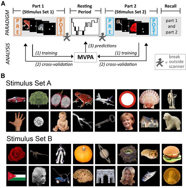
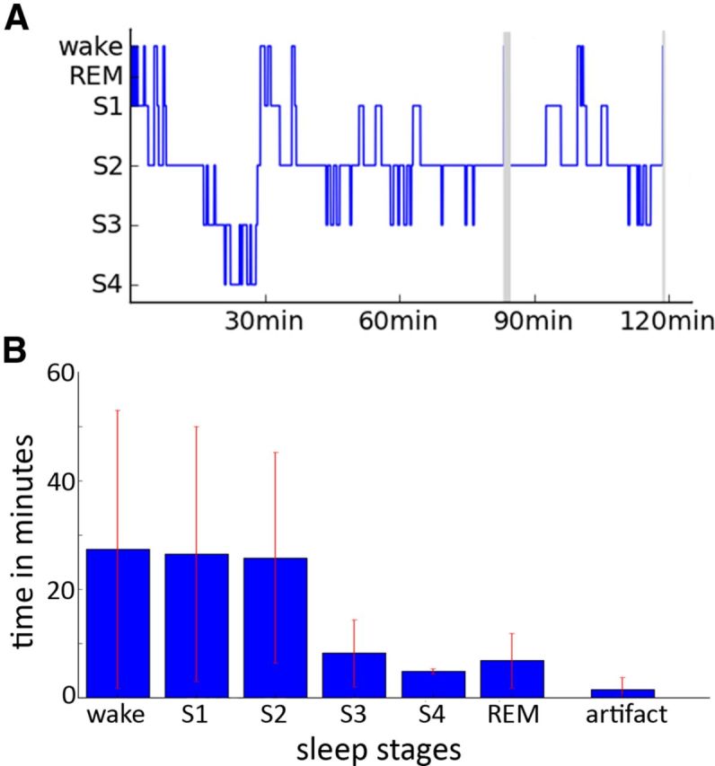
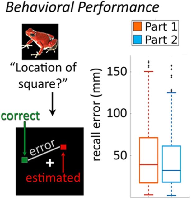
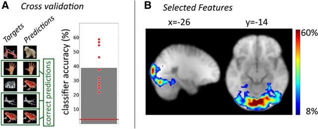
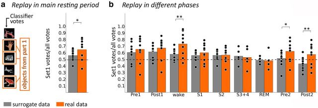
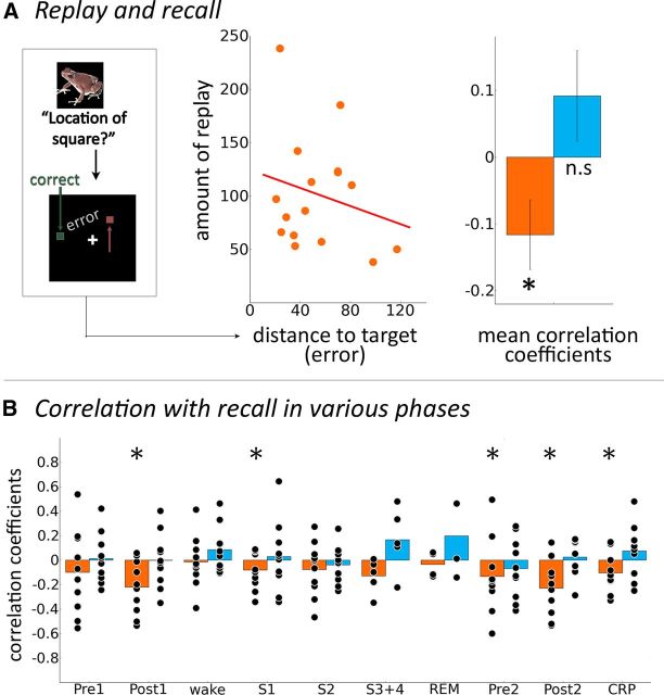
Memory consolidation transforms initially labile memory traces into more stable representations. One putative mechanism for consolidation is the reactivation of memory traces after their initial encoding during subsequent sleep or waking state. However, it is still unknown whether consolidation of individual memory contents relies on reactivation of stimulus-specific neural representations in humans. Investigating stimulus-specific representations in humans is particularly difficult, but potentially feasible using multivariate pattern classification analysis (MVPA). Here, we show in healthy human participants that stimulus-specific activation patterns can indeed be identified with MVPA, that these patterns reoccur spontaneously during postlearning resting periods and sleep, and that the frequency of reactivation predicts subsequent memory for individual items. We conducted a paired-associate learning task with items and spatial positions and extracted stimulus-specific activity patterns by MVPA in a simultaneous electroencephalography and functional magnetic resonance imaging (fMRI) study. As a first step, we investigated the amount of fMRI volumes during rest that resembled either one of the items shown before or one of the items shown as a control after the resting period. Reactivations during both awake resting state and sleep predicted subsequent memory. These data are first evidence that spontaneous reactivation of stimulus-specific activity patterns during resting state can be investigated using MVPA. They show that reactivation occurs in humans and is behaviorally relevant for stabilizing memory traces against interference. They move beyond previous studies because replay was investigated on the level of individual stimuli and because reactivations were not evoked by sensory cues but occurred spontaneously.


Figures

Figure 1.
Figure 1.
Overview of experimental paradigm. A, Subjects learned associations between 32 different stimuli (e.g., a red frog) and spatial locations that were indicated by a white square. Every object was presented 30 times followed by the corresponding location. Half of the object–location associations had to be learned in the first part of the experiment, the other half in the second part. During the main resting period between the two learning sessions, subjects slept inside an MRI scanner with simultaneous EEG. Both memory tasks were flanked by 5 min of resting state scanning (“task-adjacent resting periods”). In a memory test subsequent to the second learning task, each of the 32 objects was presented again and subjects had to indicate the position of the associated white square. B, Overview of all stimuli categories used. Note that six different exemplars were used for each stimulus type (e.g., 6 different pictures of German Chancellor Angela Merkel).
Figure 2.
Figure 2.
Sleep staging and sleep duration. A, Example of a hypnogram for one participant. Gray areas indicate phases in which sleep staging was not possible due to scanner artifacts. B, Average time spent in waking state and different sleep stages across participants. Note that for S3 and S4 only five and for rapid eye movement sleep (REM) only four participants were taken into the average as the others did not reach those stages.
Figure 3.
Figure 3.
Behavioral results from memory recall. Memory performance was measured as the distance between the correct and the indicated spatial position of the square associated with an item during the encoding phase. The box plots showing median and variance of memory performance across all recall trials and participants demonstrate relatively high intraindividual and interindividual variability.
Figure 4.
Figure 4.
Extraction of stimulus-specific representations by multivariate pattern analysis. A, Pattern classification accuracy as assessed by a cross-validation approach. Each red point indicates results from one participant. The red line indicates chance performance (3.125%). B, The classifier was trained on the 1000 most discriminative features (i.e., voxels) from each subject. The figure shows the regional distribution of features that were selected most often, which were most abundant in the occipital lobe but reached into inferior temporal cortex.
Figure 5.
Figure 5.
Spontaneous replay of stimulus-specific activity during resting state. A, Results from the main resting period between the two experiments. Bars depict the frequency with which objects from the first memory task were voted for by the classifier compared with the total amount of votes. Gray bars indicate results derived from a surrogate approach. Orange bars refer to results in the empirical data. Objects from Experiment 1 are voted for significantly more often than would be expected by chance in both empirical and surrogate data, but ratios for Experiment 1 votes to all votes are significantly higher in the empirical than in the surrogate data. B, Frequency of votes for objects from the first memory task in the task-adjacent resting periods (Pre1, Post1, Pre2, Post2) and in the different stages of the main resting period in the empirical and surrogate data. The ratio of votes for objects from the first memory task to all votes was higher in the empirical versus the surrogate classifier during the waking period, as well as during Pre2 and Post2. One star denotes puncorr < 0.05; two stars denote pcorr < 0.05.
Figure 6.
Figure 6.
Replay of stimulus-specific activity correlates with memory performance. A, Illustrative scatter plot for one participant of the relationship between the number of classifier votes for a given stimulus and the distance to target during memory recall for the respective stimulus. Right, Fisher-z-transformed correlation coefficients between stimulus-wise error during behavioral recall and stimulus-wise number of classifier votes for objects from the first memory task (orange) and the second memory task (blue). B, Fisher-z-transformed Spearman's correlation coefficients for objects from the first memory task (orange) and objects from the second memory task (blue) across different phases of the experiment, including waking state, S1, S2, SWS (S3 + 4) and rapid eye movement sleep (REM). Combined resting period (CRP) includes all resting periods following presentation of the first memory task, i.e., all resting periods where replay is possible. Fisher-z-transformed correlation coefficients were tested for consistent negativity across subjects with a one-sided t test against zero. Stars indicate phases in which there was significant consistent negativity (puncorr < 0.05; in CRP, no correction for multiple comparisons was applied). There was no consistent negativity in any phases for correlations involving objects from the second memory task.

References

    1. Axmacher N, Elger CE, Fell J. Ripples in the medial temporal lobe are relevant for human memory consolidation. Brain. 2008;131:1806–1817. doi: 10.1093/brain/awn103. - DOI - PubMed
    1. Bergmann TO, Mölle M, Diedrichs J, Born J, Siebner HR. Sleep spindle-related reactivation of category-specific cortical regions after learning face-scene associations. Neuroimage. 2012;59:2733–2742. doi: 10.1016/j.neuroimage.2011.10.036. - DOI - PubMed
    1. Bode S, Haynes JD. Decoding sequential stages of task preparation in the human brain. Neuroimage. 2009;45:606–613. doi: 10.1016/j.neuroimage.2008.11.031. - DOI - PubMed
    1. Buzsáki G. Two-stage model of memory trace formation: a role for noisy brain states. Neuroscience. 1989;31:551–570. doi: 10.1016/0306-4522(89)90423-5. - DOI - PubMed
    1. Buzsáki G. Rhythms of the brain. Oxford, New York: Oxford UP; 2006.

Publication types