. 2014 Apr 15;23(8):2157-63.
doi: 10.1093/hmg/ddt611. Epub 2013 Dec 4.

ITGB6 loss-of-function mutations cause autosomal recessive amelogenesis imperfecta

Affiliations

ITGB6 loss-of-function mutations cause autosomal recessive amelogenesis imperfecta

Shih-Kai Wang et al. Hum Mol Genet. .

Abstract

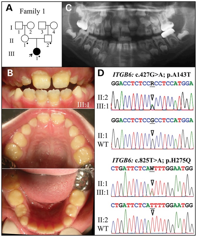
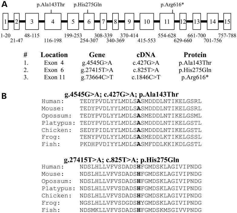
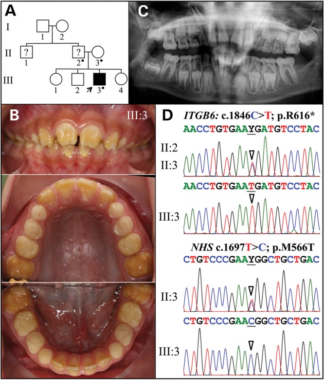
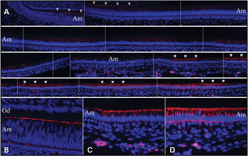
Integrins are cell-surface adhesion receptors that bind to extracellular matrices (ECM) and mediate cell-ECM interactions. Some integrins are known to play critical roles in dental enamel formation. We recruited two Hispanic families with generalized hypoplastic amelogenesis imperfecta (AI). Analysis of whole-exome sequences identified three integrin beta 6 (ITGB6) mutations responsible for their enamel malformations. The female proband of Family 1 was a compound heterozygote with an ITGB6 transition mutation in Exon 4 (g.4545G > A c.427G > A p.Ala143Thr) and an ITGB6 transversion mutation in Exon 6 (g.27415T > A c.825T > A p.His275Gln). The male proband of Family 2 was homozygous for an ITGB6 transition mutation in Exon 11 (g.73664C > T c.1846C > T p.Arg616*) and hemizygous for a transition mutation in Exon 6 of Nance-Horan Syndrome (NHS Xp22.13; g.355444T > C c.1697T > C p.Met566Thr). These are the first disease-causing ITGB6 mutations to be reported. Immunohistochemistry of mouse mandibular incisors localized ITGB6 to the distal membrane of differentiating ameloblasts and pre-ameloblasts, and then ITGB6 appeared to be internalized by secretory stage ameloblasts. ITGB6 expression was strongest in the maturation stage and its localization was associated with ameloblast modulation. Our findings demonstrate that early and late amelogenesis depend upon cell-matrix interactions. Our approach (from knockout mouse phenotype to human disease) demonstrates the power of mouse reverse genetics in mutational analysis of human genetic disorders and attests to the need for a careful dental phenotyping in large-scale knockout mouse projects.


Figures

Figure 1.
Figure 1.
Family 1. (A) Pedigree. The proband (III:1) is the only person with enamel defects in the family. A dot marks the three study participants who donated samples for DNA sequencing. (B) The proband at almost 8 years is in the mixed dentition stage with the permanent maxillary central incisors and all mandibular incisors and first molars erupted. Oral photographs of the proband show very little enamel covering dentin and signs of rapid attrition. (C) Panoramic radiograph shows no contrasting enamel layer in erupted teeth and only a thin layer of enamel in unerupted teeth, which is characteristic of hypoplastic AI. (D) Sequencing chromatograms of heterozygous ITGB6 mutations in Exon 4 (g.4545G > A; c.427G > A; p.Ala143Thr) and Exon 6 (g.27415T > A; c.825T > A; p.His275Gln). The proband was the only compound heterozygote. The father (II:2) had only the Exon 4 mutation; the mother (II:1) had only the Exon 6 mutation. The sequence variations are named relative to the ITGB6 genomic (NC_000002.11) and cDNA (NM_000888.3) reference sequences. R = G or A; W = A or T.
Figure 2.
Figure 2.
ITGB6 mutations causing AI. (A) ITGB6 gene structure diagram showing the positions of the three AI-causing mutations. The numbered boxes represent the 15 ITBG6 exons. The numbers below the exons correspond to the range of ITGB6 amino acids encoded by the exon. The bars are introns, which are not drawn to scale. The gene structure was determined by comparing the structure of the genomic reference sequence NC_000002.11 to the cDNA reference sequence NM_000888.3. (B) Multiple sequence alignments showing conservation of substitution sites in vertebrates.
Figure 3.
Figure 3.
Family 2. (A) Pedigree. The proband (III:3) is the only person with known enamel defects in the family. The proband's father and uncle were reported by the mother to have ‘yellow teeth’ but their affection status is uncertain. A dot marks the three study participants who donated samples for DNA sequencing. (B) The proband at age 8 is in the mixed dentition stage with all of the permanent maxillary and mandibular incisors and first molars erupted. Oral photographs of the proband show very little enamel covering dentin and signs of rapid attrition. (C) Panoramic radiograph shows a thin, interrupted layer of contrasting enamel in some erupted teeth and a thin layer of continuous enamel in unerupted teeth. The mandibular left second molar is missing. (D) Sequencing chromatograms of ITGB6 exon 11 (top) and NHS Exon 6 (bottom). The mother (II:3) was heterozygous for both of these sequence variations. Y = T or C.
Figure 4.
Figure 4.
ITGB6 immunohistochemistry of Day 14 mouse mandibular incisors. (A) The top left panel shows the cervical loop. Subsequent images move incisally. Single arrowheads mark ITGB6 signal along distal membrane of polarizing ameloblasts (note the varying levels of the nuclei and close proximity to the opposing sheet of odontoblasts). The early signal continues until the ameloblasts are fully polarized and the distance between the ameloblast and odontoblasts has increased. ITGB6 signal along the distal membrane diminishes and ends and ITGB6 appears to be internalized in secretory ameloblasts. Double arrowheads mark ITGB6 signal along the distal membrane in maturation stage ameloblasts. The signal along the distal membrane comes and goes, which presumably correlates with ameloblast modulations. (B) Higher magnification view of ITGB6 along the distal membrane of differentiating ameloblasts. (C) Higher magnification view of ITGB6 at the onset of enamel maturation. (D) Higher magnification view of ITGB6 late in maturation stage. Note: blue is DAPI staining of nuclei; red is ITGB6 immunofluorescence; Od, odontoblasts; Am, ameloblasts.

References

    1. Hynes R.O. Integrins: bidirectional, allosteric signaling machines. Cell. 2002;110:673–687. doi:10.1016/S0092-8674(02)00971-6. - DOI - PubMed
    1. Salmivirta K., Gullberg D., Hirsch E., Altruda F., Ekblom P. Integrin subunit expression associated with epithelial-mesenchymal interactions during murine tooth development. Dev. Dyn. 1996;205:104–113. doi:10.1002/(SICI)1097-0177(199602)205:2<104::AID-AJA2>3.0.CO;2-M. - DOI - PubMed
    1. Chen B., Goodman E., Lu Z., Bandyopadhyay A., Magraw C., He T., Raghavan S. Function of beta1 integrin in oral epithelia and tooth bud morphogenesis. J. Dent. Res. 2009;88:539–544. doi:10.1177/0022034509338008. - DOI - PMC - PubMed
    1. Yoshida T., Kumashiro Y., Iwata T., Ishihara J., Umemoto T., Shiratsuchi Y., Kawashima N., Sugiyama T., Yamato M., Okano T. Requirement of integrin beta3 for iron transportation during enamel formation. J. Dent. Res. 2012;91:1154–1159. doi:10.1177/0022034512462722. - DOI - PubMed
    1. Mohazab L., Koivisto L., Jiang G., Kytomaki L., Haapasalo M., Owen G.R., Wiebe C., Xie Y., Heikinheimo K., Yoshida T., et al. Critical role for alphavbeta6 integrin in enamel biomineralization. J. Cell Sci. 2013;126:732–744. doi:10.1242/jcs.112599. - DOI - PubMed

Publication types

MeSH terms

Supplementary concepts