. 2014 Jan;141(2):318-24.
doi: 10.1242/dev.099804. Epub 2013 Dec 4.

Involvement of Delta/Notch signaling in zebrafish adult pigment stripe patterning

Affiliations

Involvement of Delta/Notch signaling in zebrafish adult pigment stripe patterning

Hiroki Hamada et al. Development. 2014 Jan.

Erratum in

  • Development. 2014 Mar;141(6):1418

Abstract

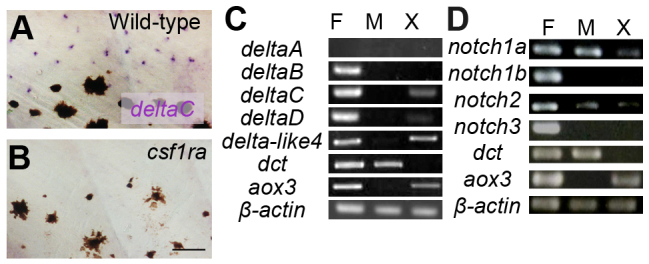
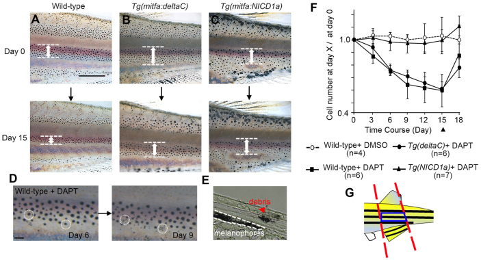
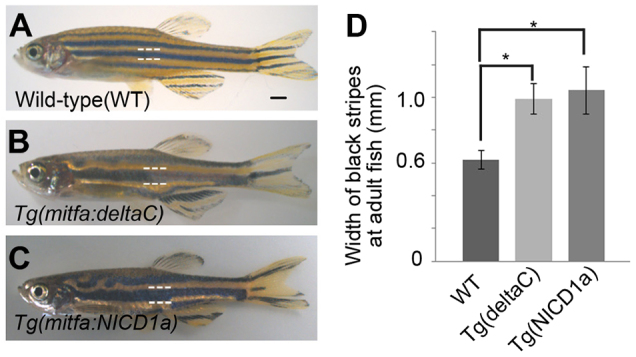
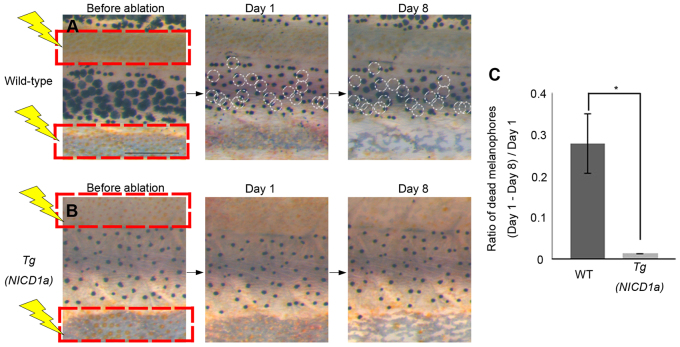
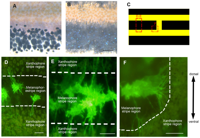
The skin pigment pattern of zebrafish is a good model system in which to study the mechanism of biological pattern formation. Although it is known that interactions between melanophores and xanthophores play a key role in the formation of adult pigment stripes, molecular mechanisms for these interactions remain largely unknown. Here, we show that Delta/Notch signaling contributes to these interactions. Ablation of xanthophores in yellow stripes induced the death of melanophores in black stripes, suggesting that melanophores require a survival signal from distant xanthophores. We found that deltaC and notch1a were expressed by xanthophores and melanophores, respectively. Moreover, inhibition of Delta/Notch signaling killed melanophores, whereas activation of Delta/Notch signaling ectopically in melanophores rescued the survival of these cells, both in the context of pharmacological inhibition of Delta/Notch signaling and after ablation of xanthophores. Finally, we showed by in vivo imaging of cell membranes that melanophores extend long projections towards xanthophores in the yellow stripes. These data suggest that Delta/Notch signaling is responsible for a survival signal provided by xanthophores to melanophores. As cellular projections can enable long-range interaction between membrane-bound ligands and their receptors, we propose that such projections, combined with direct cell-cell contacts, can substitute for the effect of a diffusible factor that would be expected by the conventional reaction-diffusion (Turing) model.

Keywords: Delta/Notch signal; Pigment pattern; Turing mechanism.


Figures

Fig. 1.
Fig. 1.
Expression analysis of Notch receptors and their ligands. (A,B) In situ hybridization for deltaC in larval fish. Presumptive xanthophores in the inter stripe are stained in wild type (A) but these cells and deltaC staining is absent in the csf1ra mutant (B). (C,D) Expression by RT-PCR of major Notch ligands (C) and Notch receptors (D). F, fins; M, melanophores; X, xanthophores. dct and aox3 are markers for melanophores and xanthophores, respectively. Scale bar: 50 μm.
Fig. 2.
Fig. 2.
Effect of Notch inhibitor on melanophores. (A-C) Altered melanophore distributions resulting from DAPT treatment over 15 days. Width of black stripes (white arrows) became narrowed in wild-type and Tg(mitfa:deltaC) fish, but remained unchanged in Tg(mitfa:NICD1a) fish. (D) Death of melanophores. Two cells (circled by dotted lines) present at day 6 (left) were lost by day 9 (right), though residual melanin-containing debris remains visible. (E) Cross-section through the skin of a DAPT-treated fish. A remaining melanophore is in the lowest layer of the skin (bounded by white dashed line). Melanin-containing debris from a dead cell was observed (red arrowhead) more superficially in the dermis. (F) Alteration of melanophore numbers (mean ± s.d.) during DAPT treatment, shown as the ratio of cells surviving at day X relative to the total number of cells at day 0. DAPT was applied at day 0 and removed at day 15 (black arrowhead). (G) Blue box shows the region in which melanophores were counted for analyses shown in F, defined as the middle stripe, delimited by anterior and posterior margins of the caudal and dorsal fins. Scale bars: 1 mm (A-C); 100 μm (D).
Fig. 3.
Fig. 3.
Ectopic expression of DeltaC and Notch-NICD alter stripes. (A-C) Expression of DeltaC (B) and the Notch1a intracellular domain (C) in melanophores resulted in fewer, wider stripes than those in wild type (A). (D) Width of black stripes in transgenic fish [wild type, n=12; Tg(mitfa:deltaC), n=12; Tg(mitfa:NICD1a), n=13]. *P<0.0001 (Student’s t-test). Error bars represent s.d. Scale bar: 1 mm.
Fig. 4.
Fig. 4.
Melanophore survival is independent of xanthophores in Tg(mitfa:NICD1a) transgenic fish. (A,B) Effects on melanophores after ablation of xanthophores is shown for wild type (A) and Tg(mitfa:NICD1a) (B). Red squares indicate the areas in which xanthophores were ablated. White circles indicate melanophores present on day 1 but lost by day 8. (C) Proportion of melanophores that died in wild-type and Tg(mitfa:NICD1a) fish (n=5, respectively). *P<0.0001 (Student’s t-test). Error bars represent s.d. Scale bar: 500 μm.
Fig. 5.
Fig. 5.
Melanophores extend long projections. (A,B) Black stripe region of wild-type fish in the normal condition (A) and after addition of yohimbine (B), which reveals projections extending towards xanthophores. (C) Regions imaged in D-F. (D-F) Fluorescent images of melanophore membranes revealed by mitfa: EGFP-CAAX expression in brassb2 mutants. (D) Three melanophores in the black stripe extend long projections towards xanthophores. (E) A cluster of melanophores, each of which exhibits long projections. (F) Melanophores adjacent to a break in the stripe extend long projections radially. Scale bars: 100 μm.

Comment in

  • Taking striping up a notch.
    Goda M, Kelsh RN, Hashimoto H. Goda M, et al. Pigment Cell Melanoma Res. 2014 Sep;27(5):688-9. doi: 10.1111/pcmr.12288. Epub 2014 Jul 15. Pigment Cell Melanoma Res. 2014. PMID: 24961581 No abstract available.

References

    1. Asai R., Taguchi E., Kume Y., Saito M., Kondo S. (1999). Zebrafish leopard gene as a component of the putative reaction-diffusion system. Mech. Dev. 89, 87–92 - PubMed
    1. Budi E. H., Patterson L. B., Parichy D. M. (2011). Post-embryonic nerve-associated precursors to adult pigment cells: genetic requirements and dynamics of morphogenesis and differentiation. PLoS Genet. 7, e1002044 - PMC - PubMed
    1. Casey P. J., Seabra M. C. (1996). Protein prenyltransferases. J. Biol. Chem. 271, 5289–5292 - PubMed
    1. Cohen M., Georgiou M., Stevenson N. L., Miodownik M., Baum B. (2010). Dynamic filopodia transmit intermittent Delta-Notch signaling to drive pattern refinement during lateral inhibition. Dev. Cell 19, 78–89 - PubMed
    1. Cooper C. D., Linbo T. H., Raible D. W. (2009). Kit and foxd3 genetically interact to regulate melanophore survival in zebrafish. Dev. Dyn. 238, 875–886 - PMC - PubMed

Publication types

MeSH terms