

Primary frozen shoulder: brief review of pathology and imaging abnormalities
- PMID: 24306579
- PMCID: PMC3929028
- DOI: 10.1007/s00776-013-0495-x
Primary frozen shoulder: brief review of pathology and imaging abnormalities
Abstract
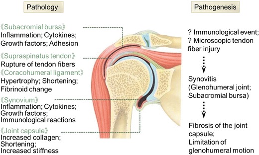
Background: Primary frozen shoulder (FS) is a painful contracture of the glenohumeral joint that arises spontaneously without an obvious preceding event. Investigation of the intra-articular and periarticular pathology would contribute to the treatment of primary FS.
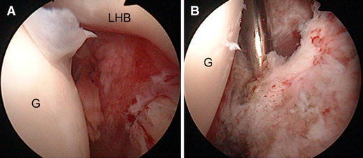
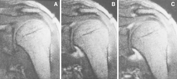
Review of literature: Many studies indicate that the main pathology is an inflammatory contracture of the shoulder joint capsule. This is associated with an increased amount of collagen, fibrotic growth factors such as transforming growth factor-beta, and inflammatory cytokines such as tumor necrosis factor-alpha and interleukins. Immune system cells such as B-lymphocytes, T-lymphocytes and macrophages are also noted. Active fibroblastic proliferation similar to that of Dupuytren's contracture is documented. Presence of inflammation in the FS synovium is supported by the synovial enhancement with dynamic magnetic resonance study in the clinical setting.
Conclusion: Primary FS shows fibrosis of the joint capsule, associated with preceding synovitis. The initiator of synovitis, however, still remains unclear. Future studies should be directed to give light to the pathogenesis of inflammation to better treat or prevent primary FS.
Figures
References
-
- Codman EA. The shoulder. New York: G. Miller & Co. Medical Publishers Inc.; 1934. p. 216–24.
-
- Duplay ES. De la périarthrite scapulohumérale et des raideurs de l’epaule qui en son la consequence. Arch Gen Med. 1872;20:513–542.
-
- Neviaser JS. Adhesive capsulitis of the shoulder. J Bone Jt Surg. 1945;27:211–222.
-
- Lundberg BJ. The frozen shoulder: clinical and radiographical observations: the effect of manipulation under general anesthesia: structure and glycosaminoglycan content of the joint capsule: local bone metabolism. Acta Orthop Scand Suppl. 1969;119:1–59. - PubMed
Publication types
MeSH terms
LinkOut - more resources
Full Text Sources
Other Literature Sources
Medical
Research Materials
