Rats with minimal hepatic encephalopathy due to portacaval shunt show differential increase of translocator protein (18 kDa) binding in different brain areas, which is not affected by chronic MAP-kinase p38 inhibition
- PMID: 24307181
- PMCID: PMC4087148
- DOI: 10.1007/s11011-013-9461-8
Rats with minimal hepatic encephalopathy due to portacaval shunt show differential increase of translocator protein (18 kDa) binding in different brain areas, which is not affected by chronic MAP-kinase p38 inhibition
Abstract
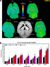
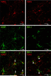
Neuroinflammation plays a main role in neurological deficits in rats with minimal hepatic encephalopathy (MHE) due to portacaval shunt (PCS). Treating PCS rats with SB239063, an inhibitor of MAP-kinase-p38, reduces microglial activation and brain inflammatory markers and restores cognitive and motor function. The translocator protein-(18-kDa) (TSPO) is considered a biomarker of neuroinflammation. TSPO is increased in brain of PCS rats and of cirrhotic patients that died in hepatic coma. Rats with MHE show strong microglial activation in cerebellum and milder in other areas when assessed by MHC-II immunohistochemistry. This work aims were assessing: 1) whether binding of TSPO ligands is selectively increased in cerebellum in PCS rats; 2) whether treatment with SB239063 reduces binding of TSPO ligands in PCS rats; 3) which cell type (microglia, astrocytes) increases TSPO expression. Quantitative autoradiography was used to assess TSPO-selective (3)H-(R)-PK11195 binding to different brain areas. TSPO expression increased differentially in PCS rats, reaching mild expression in striatum or thalamus and very high levels in cerebellum. TSPO was expressed in astrocytes and microglia. Treatment with SB239063 did not reduces (3)[H]-PK11195 binding in PCS rats. SB239063 reduces microglial activation and levels of inflammatory markers, but not binding of TSPO ligands. This indicates that SB239063-induced neuroinflammation reduction in PCS rats is not mediated by effects on TSPO. Also, enhanced TSPO expression is not always associated with cognitive or motor deficits. If enhanced TSPO expression plays a role in mechanisms leading to neurological alterations in MHE, SB239063 would interfere these mechanisms at a later step.
Figures


Similar articles
-
p38 MAP kinase is a therapeutic target for hepatic encephalopathy in rats with portacaval shunts.Gut. 2011 Nov;60(11):1572-9. doi: 10.1136/gut.2010.236083. Epub 2011 Jun 2. Gut. 2011. PMID: 21636647
-
Sildenafil reduces neuroinflammation and restores spatial learning in rats with hepatic encephalopathy: underlying mechanisms.J Neuroinflammation. 2015 Oct 29;12:195. doi: 10.1186/s12974-015-0420-7. J Neuroinflammation. 2015. PMID: 26511444 Free PMC article.
-
Neuroinflammation increases GABAergic tone and impairs cognitive and motor function in hyperammonemia by increasing GAT-3 membrane expression. Reversal by sulforaphane by promoting M2 polarization of microglia.J Neuroinflammation. 2016 Apr 18;13(1):83. doi: 10.1186/s12974-016-0549-z. J Neuroinflammation. 2016. PMID: 27090509 Free PMC article.
-
Peripheral inflammation induces neuroinflammation that alters neurotransmission and cognitive and motor function in hepatic encephalopathy: Underlying mechanisms and therapeutic implications.Acta Physiol (Oxf). 2019 Jun;226(2):e13270. doi: 10.1111/apha.13270. Epub 2019 Mar 22. Acta Physiol (Oxf). 2019. PMID: 30830722 Review.
-
Neurosteroids in hepatic encephalopathy: Novel insights and new therapeutic opportunities.J Steroid Biochem Mol Biol. 2016 Jun;160:94-7. doi: 10.1016/j.jsbmb.2015.11.006. Epub 2015 Nov 14. J Steroid Biochem Mol Biol. 2016. PMID: 26589093 Review.
Cited by
-
Strategies for Improving Photodynamic Therapy Through Pharmacological Modulation of the Immediate Early Stress Response.Methods Mol Biol. 2022;2451:405-480. doi: 10.1007/978-1-0716-2099-1_20. Methods Mol Biol. 2022. PMID: 35505025 Review.
-
[18F]PBR146 and [18F]DPA-714 in vivo Imaging of Neuroinflammation in Chronic Hepatic Encephalopathy Rats.Front Neurosci. 2021 Aug 16;15:678144. doi: 10.3389/fnins.2021.678144. eCollection 2021. Front Neurosci. 2021. PMID: 34483820 Free PMC article.
-
Recent advances in hepatic encephalopathy.F1000Res. 2017 Sep 4;6:1637. doi: 10.12688/f1000research.11938.1. eCollection 2017. F1000Res. 2017. PMID: 29026534 Free PMC article. Review.
-
Acetyl-lysine binding site of bromodomain-containing protein 4 (BRD4) interacts with diverse kinase inhibitors.ACS Chem Biol. 2014 May 16;9(5):1160-71. doi: 10.1021/cb500072z. Epub 2014 Mar 13. ACS Chem Biol. 2014. PMID: 24568369 Free PMC article.
-
The neurogliovascular unit in hepatic encephalopathy.JHEP Rep. 2021 Aug 11;3(5):100352. doi: 10.1016/j.jhepr.2021.100352. eCollection 2021 Oct. JHEP Rep. 2021. PMID: 34611619 Free PMC article. Review.
References
-
- Agusti A, Cauli O, Rodrigo R, Llansola M, Hernandez-Rabaza V, Felipo V. p38 MAP kinase is a therapeutic target for hepatic encephalopathy in rats with portacaval shunts. Gut. 2011;60:1572–1579. - PubMed
-
- Ahboucha S, Butterworth RF. The neurosteroid system: an emerging therapeutic target for hepatic encephalopathy. Metab Brain Dis. 2007;22(3–4):291–308. - PubMed
-
- Ahboucha S, Butterworth RF. The neurosteroid system: implication in the pathophysiology of hepatic encephalopathy. Neurochem Int. 2008;52(4–5):575–587. - PubMed
-
- Ahboucha S, Pomier-Layrargues G, Mamer O, Butterworth RF. Increased levels of pregnenolone and its neuroactive metabolite allopregnanolone in autopsied brain tissue from cirrhotic patients who died in hepatic coma. Neurochem Int. 2006;49:372–378. - PubMed
-
- Amodio P, Montagnese S, Gatta A, Morgan MY. Characteristics of minimal hepatic encephalopathy. Metab Brain Dis. 2004;19:253–267. - PubMed
Publication types
MeSH terms
Substances
Grants and funding
LinkOut - more resources
Full Text Sources
Other Literature Sources
Research Materials

