. 2014 Jul;19(4):493-505.
doi: 10.1007/s12192-013-0475-2. Epub 2013 Dec 5.

Prognostic implication of HSPA (HSP70) in breast cancer patients treated with neoadjuvant anthracycline-based chemotherapy

Affiliations

Prognostic implication of HSPA (HSP70) in breast cancer patients treated with neoadjuvant anthracycline-based chemotherapy

Silvina B Nadin et al. Cell Stress Chaperones. 2014 Jul.

Abstract

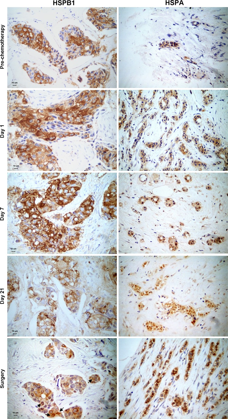
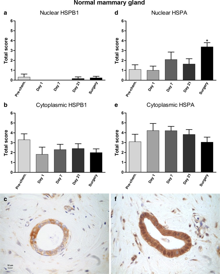
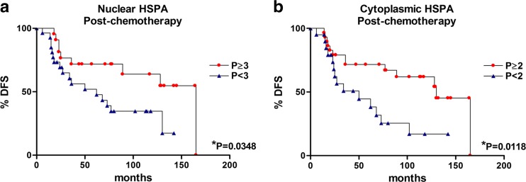
Neoadjuvant chemotherapy is used in patients with locally advanced breast cancer to reduce tumor size before surgery. Unfortunately, resistance to chemotherapy may arise from a variety of mechanisms. Heat shock proteins (HSPs), which are highly expressed in mammary tumor cells, have been implicated in anticancer drug resistance. In spite of the widely described value of HSPs as molecular markers in cancer, their implications in breast tumors treated with anthracycline-based neoadjuvant chemotherapy has been poorly explored. In this study, we have evaluated, by immunohistochemistry, the expression of HSP27 (HSPB1) and HSP70 (HSPA) in serial biopsies from locally advanced breast cancer patients (n = 60) treated with doxorubicin (DOX)- or epirubicin (EPI)-based monochemotherapy. Serial biopsies were taken at days 1, 3, 7, and 21, and compared with prechemotherapy and surgical biopsies. After surgery, the patients received additional chemotherapy with cyclophosphamide, methotrexate, and 5-fluorouracil. High nuclear HSPB1 and HSPA expressions were found in invasive cells after DOX/EPI administration (P < 0.001), but the drug did not affect the cytoplasmic expression of the HSPs. Infiltrating lymphocytes showed high nuclear HSPA (P < 0.01) levels at postchemotherapy. No correlations were found between HSPs expression and the clinical and pathological response to neoadjuvant therapy. However, in postchemotherapy biopsies, high nuclear (>31 % of the cells) and cytoplasmic HSPA expressions (>11 % of the tumor cells) were associated with better DFS (P = 0.0348 and P = 0.0118, respectively). We conclude that HSPA expression may be a useful prognostic marker in breast cancer patients treated with neoadjuvant DOX/EPI chemotherapy indicating the need to change the administered drugs after surgery for overcoming drug resistance.


Figures

Fig. 1
Fig. 1
Expression of HSPB1 and HSPA in invasive tumor cells before, during (days 1, 7, and 21), and after neoadjuvant chemotherapy. HSPB1 was expressed mainly in the cytoplasm of tumor cells. Observe some HSPB1 positive nuclei in the surgery specimen (arrows). Note the cytoplasmic expression of HSPA before DOX administration and the predominant nuclear expression shortly after chemotherapy. The positive immunoreactivity appears as brown deposits, and the slides were lightly counterstained with hematoxylin to reveal nuclei. A scale bar (10 μm) was included in the left microphotographs
Fig. 2
Fig. 2
HSPB1 and HSPA expression in invasive carcinomas before and after chemotherapy. a, b Nuclear HSPB1 and HSPA expression, respectively. c, d Cytoplasmic HSPB1 and HSPA expression, respectively. e, f Samples showing HSPB1 and HSPA, respectively. Note the nuclear and cytoplasmic expression of both HSPs and cell membrane immunoreactions (arrows). A 10 μm scale bar was included in the microphotographs. Total score represents the intensity and proportion of the positive immunostainings (see M&M section). Bars represent mean ± SEM. ***P < 0.001
Fig. 3
Fig. 3
Expressions of HSPB1 and HSPA in normal mammary tissue before and after neoadjuvant chemotherapy. a, d Nuclear HSPB1 and HSPA expressions, respectively. b, e Cytoplasmic HSPB1and HSPA expressions, respectively. c, f Images of normal breast ducts from postchemotherapy biopsies with positive staining for HSPB1 and HSPA, respectively. Note the accumulation of HSPA in the nucleus of the ductal epithelial cells. Bars represent mean ± SEM. A 10 μm scale bar was included in one of the microphotographs. Total score represents the intensity and proportion of the positive immunostaining.*P < 0.05
Fig. 4
Fig. 4
Tumor-infiltrating lymphocytes before, during (days 7 and 21), and after neoadjuvant anthracycline-based chemotherapy. a Intratumoral lymphocytes mean ± SEM. b Peritumoral lymphocytes mean ± SEM. c, d Photomicrographs taken from prechemotherapy biopsy and surgery specimen from one breast cancer patient, respectively. A 10 μm scale bar was included in one of the microphotographs. *P < 0.05; **P < 0.01
Fig. 5
Fig. 5
HSPA expression in lymphocytes and fibroblasts in breast biopsies before and after neoadjuvant chemotherapy. a, d Images taken from a postchemotherapy biopsy. A 10 μm scale bar was included in one of the microphotographs. Note the positive HSPA nuclear staining. b, e Nuclear HSPA expression in lymphocytes and fibroblasts, respectively. c, f Cytoplasmic HSPA expression in lymphocytes and fibroblasts, respectively. Total score represents the intensity and proportion of the positive immunostaining. Bars represent mean ± SEM. *P < 0.05; **P < 0.01
Fig. 6
Fig. 6
Disease-free survival (DFS) curves in function of nuclear and cytoplasmic HSPA expression. High HSPA nuclear expression (proportion ≥3) and high HSPA cytoplasmic expression (proportion ≥2) at postchemotherapy correlated with DFS (a and b, respectively). The exact P value is indicated in each figure. *P < 0.05

Similar articles

Cited by

References

    1. Almeida MB, do Nascimento JL, Herculano AM, Crespo-López ME. Molecular chaperones: toward new therapeutic tools. Biomed Pharmacother. 2011;65:239–243. doi: 10.1016/j.biopha.2011.04.025. - DOI - PubMed
    1. Apetoh L, Ghiringhelli F, Tesniere A, Obeid M, Ortiz C, Criollo A, Mignot G, Maiuri MC, Ullrich E, Saulnier P, Yang H, Amigorena S, Ryffel B, Barrat FJ, Saftig P, Levi F, Lidereau R, Nogues C, Mira JP, Chompret A, Joulin V, Clavel-Chapelon F, Bourhis J, André F, Delaloge S, Tursz T, Kroemer G, Zitvogel L. Toll-like receptor 4-dependent contribution of the immune system to anticancer chemotherapy and radiotherapy. Nat Med. 2007;13:1050–1059. doi: 10.1038/nm1622. - DOI - PubMed
    1. Apetoh L, Ghiringhelli F, Tesniere A, Obeid M, Mignot G, Ullrich E, Kroemer G, Zitvogel L. Cancer is not just a disease of a tissue: it is a host disease. How to reactivate host defense against tumors using conventional therapies of cancer? Ann Endocrinol (Paris) 2008;69:151–152. doi: 10.1016/j.ando.2008.02.016. - DOI - PubMed
    1. Apetoh L, Tesniere A, Ghiringhelli F, Kroemer G, Zitvogel L. Molecular interactions between dying tumor cells and the innate immune system determine the efficacy of conventional anticancer therapies. Cancer Res. 2008;68:4026–4030. doi: 10.1158/0008-5472.CAN-08-0427. - DOI - PubMed
    1. Binaschi M, Bigioni M, Cipollone A, Rossi C, Goso C, Maggi CA, Capranico G, Animati F. Anthracyclines: selected new developments. Curr Med Chem Anticancer Agents. 2001;1:113–130. doi: 10.2174/1568011013354723. - DOI - PubMed

Publication types

MeSH terms