. 2014 Feb;88(4):1990-9.
doi: 10.1128/JVI.03312-13. Epub 2013 Dec 4.

Inhibition of hepatitis C virus production by aptamers against the core protein

Affiliations

Inhibition of hepatitis C virus production by aptamers against the core protein

Shali Shi et al. J Virol. 2014 Feb.

Abstract

Hepatitis C virus (HCV) core protein is essential for virus assembly. HCV core protein was expressed and purified. Aptamers against core protein were raised through the selective evolution of ligands by the exponential enrichment approach. Detection of HCV infection by core aptamers and the antiviral activities of aptamers were characterized. The mechanism of their anti-HCV activity was determined. The data showed that selected aptamers against core specifically recognize the recombinant core protein but also can detect serum samples from hepatitis C patients. Aptamers have no effect on HCV RNA replication in the infectious cell culture system. However, the aptamers inhibit the production of infectious virus particles. Beta interferon (IFN-β) and interferon-stimulated genes (ISGs) are not induced in virally infected hepatocytes by aptamers. Domains I and II of core protein are involved in the inhibition of infectious virus production by the aptamers. V31A within core is the major resistance mutation identified. Further study shows that the aptamers disrupt the localization of core with lipid droplets and NS5A and perturb the association of core protein with viral RNA. The data suggest that aptamers against HCV core protein inhibit infectious virus production by disrupting the localization of core with lipid droplets and NS5A and preventing the association of core protein with viral RNA. The aptamers for core protein may be used to understand the mechanisms of virus assembly. Core-specific aptamers may hold promise for development as early diagnostic reagents and potential therapeutic agents for chronic hepatitis C.


Figures
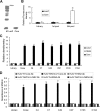
FIG 1
FIG 1
Selection of aptamers against HCV core protein and binding affinity of the aptamers. (A) His-tagged core was expressed by IPTG (isopropyl-β-d-thiogalactopyranoside) induction in E. coli BL21(DE3). The core protein and control protein LacZ were separated on SDS-PAGE gel and stained using mouse anti-His monoclonal antibody via Western blotting. (B) FITC-labeled DNA pools from library round 1 and round 8 were incubated with agarose beads conjugated with HCV core or control protein LacZ in binding buffer. The density of the fluorescence was measured and normalized to library. (C) Binding affinity of core aptamers. Each biotin-labeled aptamer was added to the microtiter plate, and an ELONA was performed. Purified His-tagged core, NS5A, or control protein LacZ was added to the plates. Mouse monoclonal anti-His antibody and HRP-conjugated goat anti-mouse IgG were used as primary and secondary antibodies, respectively. Color development was performed, and the plates were read with an ELISA reader. The absorbance of each sample was measured at 450 nm and normalized to library. The data represent the averages of five different experiments. **, P < 0.01 versus library. (D) Binding affinity of core aptamers to core protein from lysates of HCV-infected hepatocytes. Biotin-labeled library, Cnew, C4, C7, C42, C97, C103, or C104 was added to the microtiter plate previously coated with streptavidin. Lysates of HCV-infected Huh7.5 or noninfected Huh7.5 cells were added to the plates. After washing, mouse anti-HCV core, NS2, or NS5A monoclonal antibody was added and incubated at 37°C for 1 h. HRP-conjugated goat anti-mouse IgG was added to the plates. The data were obtained as described for panel C and represent the means of 3 different experiments. **, P < 0.01 versus library.
FIG 2
FIG 2
Detection of HCV core in serum samples from HCV-infected patients by core aptamer. (A) A standard curve for core detected by ELONA or ELISA is shown. Both ELONA and ELISA were performed to determine the detection limitation of HCV core protein using C7 aptamer. The absorbance of each sample was measured at 450 nm. A standard curve was created by plotting the mean absorbance for each standard concentration (y axis) against the core protein concentration (x axis). Mean values and standard deviations for three independent experiments performed in triplicate are shown. (B) Detection of core in the serum samples from hepatitis C patients by core aptamer and HCV viral titer is proportional to core protein concentration in the samples. Streptavidin-precoated microtiter plates were coated with biotin-labeled core aptamer C7. The serum samples from HCV-infected patients, HBV-infected patients, or healthy donors were added into the plates. Color development was performed, and the absorbance of each sample was measured at 450 nm. HCV RNA copy numbers in the sera measured by quantitative real-time PCR were plotted against core concentration in serum samples.
FIG 3
FIG 3
Inhibition of infectious virus production by aptamers against core. (A and B) Effects of core aptamers on the intracellular (A) or extracellular (B) viral RNA level in hepatocytes. JFH1 virus suspension at a multiplicity of infection (MOI) of 0.1 was used to infect Huh7.5 cells for 3 days. The cells were treated by aptamer or library for 72 h. Intracellular or extracellular HCV RNA was measured by real-time PCR. The data represent the means of 3 different experiments. (C) Uptake efficiency of core-specific aptamers by HCV-infected hepatocytes. HCV-infected Huh7.5 cells were inoculated with different doses of Cy5-labeled aptamers. At the indicated time points, the virus-containing supernatant and the cells were collected separately. The cells were suspended in 1 ml fresh DMEM and then were subjected to four freeze-and-thaw cycles to collect the intracellular Cy5-labeled aptamers. The intracellular and extracellular supernatants were transported to the black 96-well plate at 100-μl volume per well. Then, the extracellular and intracellular Cy5 signal absorbances at 647 nm were measured by microplate reader. The percentages of the intracellular and the extracellular efficiencies were relative to the positive ones, which were the fresh culture media to which were added the Cy5-labeled aptamers at the same concentration. The data represent three independent experiments performed in triplicate. (D) Colocalization of core-specific aptamers with HCV core protein in the HCV-infected hepatocytes. HCV-infected Huh7.5 cells were treated with Cy5-labeled aptamers for 24 h. The cells were fixed with ice-cold acetone for 10 min at −20°C. The cells were washed with PBS, blocked with 1:50 goat serum for 30 min at room temperature, and then incubated for 1 h with mouse monoclonal anticore antibody. The cells were stained with Texas Red-labeled goat anti-mouse antibody for 45 min at room temperature. The nuclei were counterstained with DAPI. Fluorescent images were obtained under a fluorescence microscope. (E and F) Effects of core aptamers on the extracellular (E) or intracellular (F) core protein level in HCV-infected hepatocytes. The cells were treated as described for panel A. Core protein in cell culture supernatant or inside the cells was measured by core protein-specific ELISA. Means and standard deviations of three independent experiments performed at least in triplicate are shown. (G) Titration of infectious virus particles produced in the presence of core aptamers by focus-forming unit assay. The cells were treated with 10 nM or 100 nM aptamer or library for 72 h. The extracellular and intracellular virus particles were harvested 72 h postinfection, and titers were determined by FFU assay on naive Huh7.5 cells. The data represent the averages of three different experiments. (H) Effects of core aptamer on viability of HCV-infected Huh7.5 cells. HCV-infected Huh7.5 cells were treated by aptamers for 72 h. The effect of aptamer on viability of the cells was measured by MTS assay. The data were normalized with the control and represent means of three independent experiments. *, P < 0.05 versus library-treated cells. (I) Core-specific aptamers do not affect hepatitis B viral DNA replication. HepG2.2.15 cells were inoculated with different doses of aptamers for 72 h. Intracellular HBV DNA was detected with real-time PCR and normalized with GAPDH. The data represent three independent experiments.
FIG 4
FIG 4
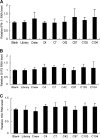
Core aptamers do not stimulate innate immunity in HCV-infected human hepatocytes. A JFH1 virus suspension at an MOI of 0.1 was used to infect Huh7.5 cells for 3 days. The cells were treated by 100 nM aptamer or library. Total cellular RNA was isolated. The levels of IFN-β (A), G1P3 (B), and 1-8U (C) mRNA were examined by real-time PCR and normalized with GAPDH. The data represent means of three different experiments.
FIG 5
FIG 5
Domain I and domain II of core protein are involved in the inhibition of infectious virus production by core-specific aptamers. (A) Confirmation of the expression of different domains of core protein by Western blotting. Different domains of core gene were cloned into expression vector separately. Different truncated versions of core protein were expressed and purified. The purified protein was detected by Western blotting. (B) Binding affinity of core aptamers to domain I or domain II of core protein. Biotin-labeled core aptamer or library was added to the microtiter plate previously coated with streptavidin, and ELONA was performed. Purified different truncated versions of core protein were added to the plates. LacZ protein was used as a control. ELONA was performed as described for Fig. 1C. Results are the averages of three independent experiments performed in triplicate. *, P < 0.05; **, P < 0.01. (C) Domains I and II of core protein are involved in the inhibition of infectious virus production by aptamers. A JFH1 virus suspension at an MOI of 0.1 was used to infect Huh7.5 cells for 3 days, and the cells were transfected with plasmids containing different truncated versions of core. The cells were then treated by 100 nM aptamer or library for 72 h. The supernatants and intracellular virus particles were harvested, and titers were determined by FFU assay. The infectivity titers in the supernatant or inside the cells with aptamer treatment were normalized to the library-treated group, and the data represent 3 different infections. *, P < 0.05; **, P < 0.01 versus vector-transfected cells.
FIG 6
FIG 6
Core aptamers disrupt the localization of core with lipid droplets and block the interaction between core and NS5A protein. (A) Subcellular localization of core protein with lipid droplets in HCV-infected hepatocytes with library or aptamer treatment. Seventy-two hours after library or core aptamer treatment, the cells were fixed with ice-cold acetone and stained with antibody against core (red). Lipid droplets were detected with the Bodipy 493/503 dye (green), followed by nuclear counterstaining with DAPI. An identical setting was maintained for image capture. Representative images are shown. (B) Effects of aptamer on the interaction between core and NS5A protein. Protein was isolated from the HCV-infected Huh7.5 cells with aptamer or library treatment and immunoprecipitated with antibodies against NS5A or mouse IgG conjugated with agarose beads, respectively. The proteins binding to the beads were boiled and subjected to SDS-PAGE. The proteins were transferred onto PVDF membrane and allowed to react with primary and secondary antibodies. Core protein was detected with Western blotting and quantified by densitometry in comparison with NS5A protein in the immunoprecipitates. Protein from HCV-infected Huh7.5 with aptamer or library treatment used for equal loading is shown as the input. The input core or NS5A protein was detected with Western blotting and quantified by densitometry. The data represent the means of 3 independent experiments. *, P < 0.05 versus library-treated cells.
FIG 7
FIG 7
Core-specific aptamers block the association of core protein with viral RNA. IP real-time PCR for HCV RNA in virus-infected hepatocytes with aptamer or library treatment was performed to examine the association between core protein and viral RNA. A JFH1 virus suspension at an MOI of 0.1 was used to infect Huh7.5 cells for 3 days. The cells were treated by 100 nM each aptamer or library for 72 h. After IP with mouse monoclonal anticore antibody or mouse control IgG, immunoprecipitates were eluted. RNA in the immunocomplexes was isolated, and real-time PCR was carried out as described in Materials and Methods. The data represent the means of three different experiments. *, P < 0.05; **, P < 0.01; ***, P < 0.001 versus library.
FIG 8
FIG 8
The V31A substitution in core is the major selective resistance mutation identified. Selection of resistance-conferring mutations was performed. Huh7.5 cells were infected with medium of Huh7.5 cells transfected with RNA from wild-type (WT) or the selected V31A viral clone. The effect of C4 on the intracellular and extracellular infectious FFU of the wild-type and the selected V31A virus was examined as described for Fig. 3E. The infectivity titers inside the cells (A) and the supernatant (B) with C4 treatment were normalized to the control group. The data represent 3 independent infections. *, P < 0.05; **, P < 0.01 versus control cells.

Similar articles

Cited by

References

    1. Lavanchy D. 2009. The global burden of hepatitis C. Liver Int. 29(Suppl 1):74–81. 10.1111/j.1478-3231.2008.01934.x - DOI - PubMed
    1. Gaudy C, Thevenas C, Tichet J, Mariotte N, Goudeau A, Dubois F. 2005. Usefulness of the hepatitis C virus core antigen assay for screening of a population undergoing routine medical checkup. J. Clin. Microbiol. 43:1722–1726. 10.1128/JCM.43.4.1722-1726.2005 - DOI - PMC - PubMed
    1. Bouvier-Alias M, Patel K, Dahari H, Beaucourt S, Larderie P, Blatt L, Hezode C, Picchio G, Dhumeaux D, Neumann AU, McHutchison JG, Pawlotsky JM. 2002. Clinical utility of total HCV core antigen quantification: a new indirect marker of HCV replication. Hepatology 36:211–218. 10.1053/jhep.2002.34130 - DOI - PubMed
    1. Rice CM. 2011. New insights into HCV replication: potential antiviral targets. Top. Antivir. Med. 19:117–120 http://www.iasusa.org/sites/default/files/tam/19-3-117.pdf - PMC - PubMed
    1. Yi M, Ma Y, Yates J, Lemon SM. 2009. Trans-complementation of an NS2 defect in a late step in hepatitis C virus (HCV) particle assembly and maturation. PLoS Pathog. 5:e1000403. 10.1371/journal.ppat.1000403 - DOI - PMC - PubMed

Publication types

MeSH terms