. 2014 Feb;88(4):1961-71.
doi: 10.1128/JVI.02467-13. Epub 2013 Dec 4.

Interactions between herpesvirus entry mediator (TNFRSF14) and latency-associated transcript during herpes simplex virus 1 latency

Affiliations

Interactions between herpesvirus entry mediator (TNFRSF14) and latency-associated transcript during herpes simplex virus 1 latency

Sariah J Allen et al. J Virol. 2014 Feb.

Abstract

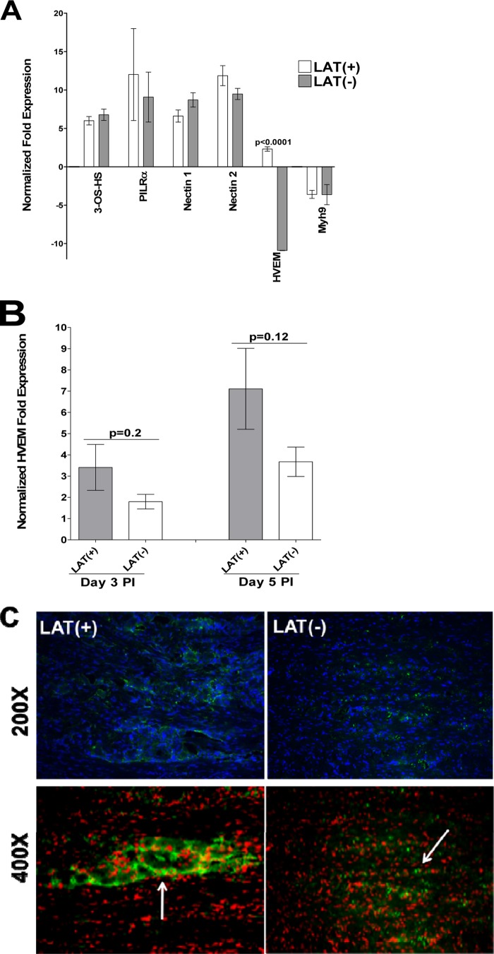
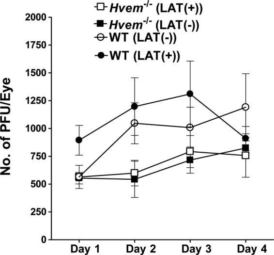
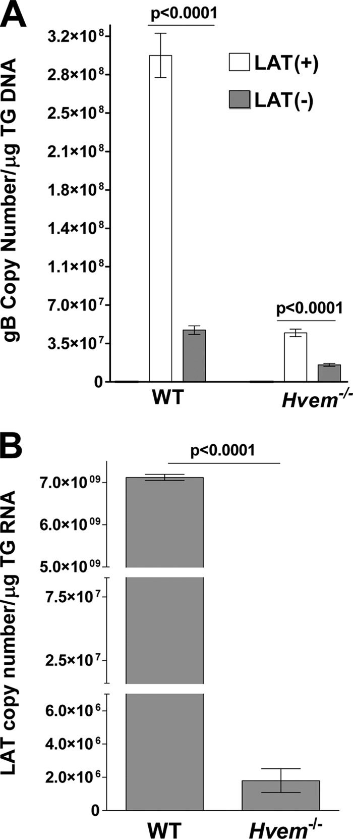
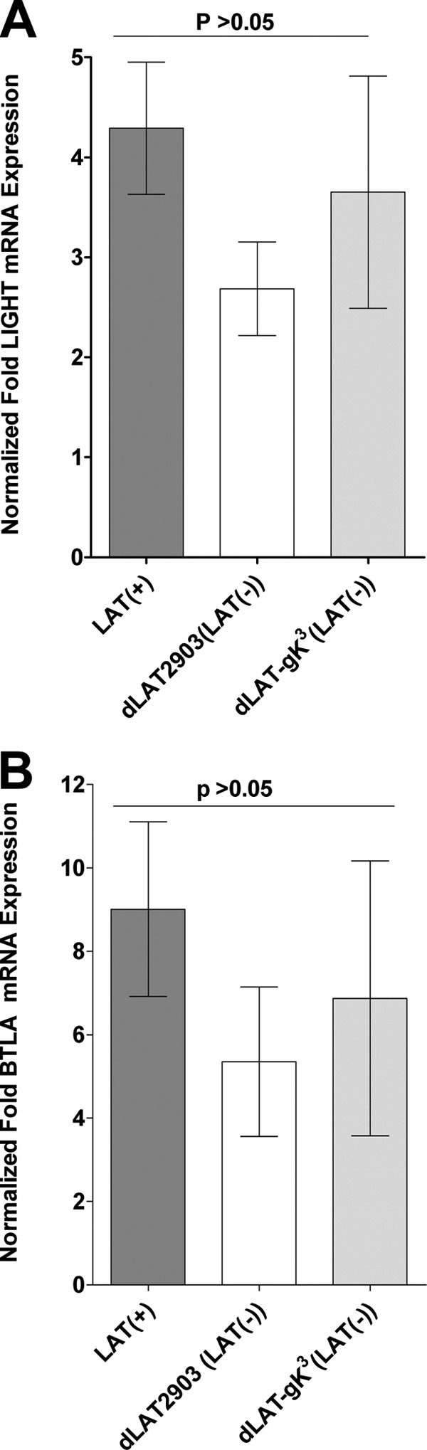
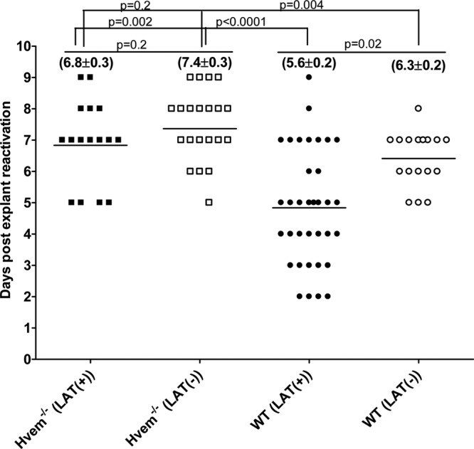
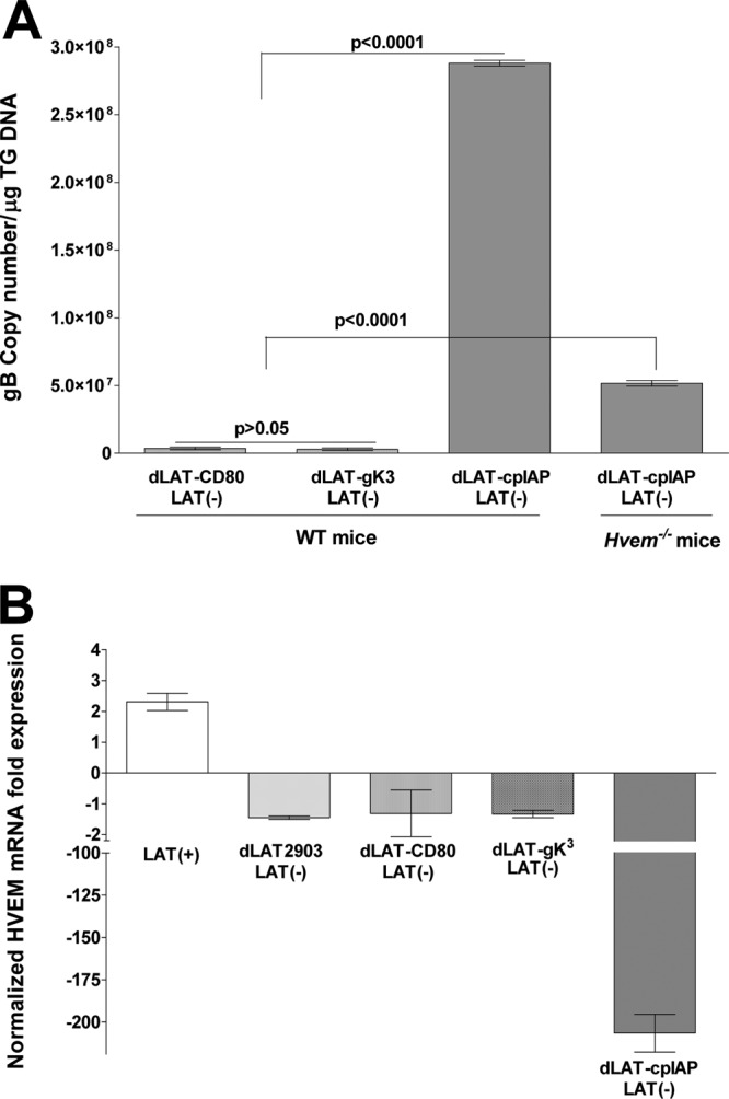
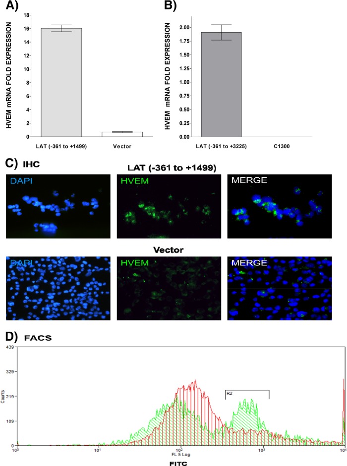
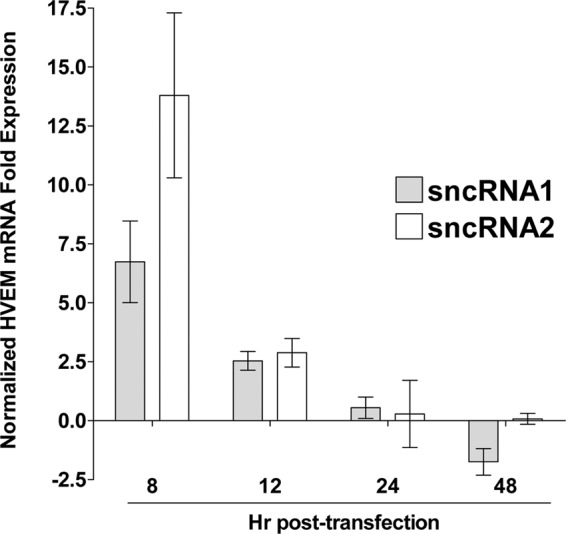
Herpesvirus entry mediator (HVEM) is one of several cell surface proteins herpes simplex virus (HSV) uses for attachment/entry. HVEM regulates cellular immune responses and can also increase cell survival. Interestingly, latency-associated transcript (LAT), the only viral gene consistently expressed during neuronal latency, enhances latency and reactivation by promoting cell survival and by helping the virus evade the host immune response. However, the mechanisms of these LAT activities are not well understood. We show here for the first time that one mechanism by which LAT enhances latency and reactivation appears to be by upregulating HVEM expression. HSV-1 latency/reactivation was significantly reduced in Hvem(-/-) mice, indicating that HVEM plays a significant role in HSV-1 latency/reactivation. Furthermore, LAT upregulated HVEM expression during latency in vivo and also when expressed in vitro in the absence of other viral factors. This study suggests a mechanism whereby LAT upregulates HVEM expression potentially through binding of two LAT small noncoding RNAs to the HVEM promoter and that the increased HVEM then leads to downregulation of immune responses in the latent microenvironment and increased survival of latently infected cells. Thus, one of the mechanisms by which LAT enhances latency/reactivation appears to be through increasing expression of HVEM.


Figures

FIG 1
FIG 1
Effect of LAT on HVEM expression in TG of infected mice. (A) Effect of LAT on expression of HSV-1 receptors in latently infected mice. C57BL/6 mice were ocularly infected with HSV-1 strain McKrae [LAT(+)] or dLAT2903 [LAT(−)]; the TG from surviving mice were isolated individually on day 30 postinfection, and quantitative RT-PCR was performed using total RNA. Nectin-1, nectin-2, HVEM, PILRα, NMHC-IIA, and 3-O-sulfated heparin sulfate (3-OS-HS) expression in naive mice was used to estimate the relative expression of each transcript in TG. GAPDH expression was used to normalize the relative expression of each transcript in TG of latently infected mice. Each bar represents the mean ± standard error of the mean from 20 TG. (B) Expression of HVEM in TG of WT infected mice during primary infection. C57BL/6 mice were infected ocularly with McKrae [LAT(+)] or dLAT2903 [LAT(−)], and expression of HVEM in TG was determined on days 3 and 5 p.i. as described above. GAPDH expression was used to normalize the relative expression of each transcript in TG of latently infected mice. Each point represents the mean ± standard error of the mean from 10 TG. (C) Upregulation of HVEM in TG of mice infected with LAT(+) virus. C57BL/6 mice were infected as described above. At 30 days p.i., TG from mice latently infected as indicated were isolated and stained with HVEM antibody as described in Materials and Methods. Nuclei are stained with DAPI (blue), and HVEM is stained in green. With LAT(+) virus infection, staining appears mostly at the surface of large cells (arrow), likely neurons. With LAT(−) virus infection, staining is mostly of small nonneuronal-like cells (arrow). Magnifications are indicated at the right of the panels.
FIG 2
FIG 2
Virus titers in WT and HVEM−/− eyes during primary ocular infection. WT C57BL/6 and C57BL/6 HVEM−/− mice were infected ocularly with LAT(+) or LAT(−) virus, and the amount of infectious HSV-1 in tear films was determined daily by standard plaque assays as described in Materials and Methods. For each time point, the virus titer (y axis) represents the average of the titers from 20 eyes ± standard error of the mean.
FIG 3
FIG 3
Effect of LAT and HVEM on HSV-1 latency and reactivation in TG of latently infected mice. WT and HVEM−/− mice were ocularly infected with HSV-1 strain McKrae [LAT(+)] or dLAT2903 [LAT(−)] as described in the legend of Fig. 1. On day 30 p.i., TG were harvested from the latently infected surviving mice. Quantitative PCR and RT-PCR were performed on each individual mouse TG. In each experiment, an estimated relative copy number of gB or LAT was calculated using a standard curve generated from pGem-gB1 or pGEM-5317, respectively. Briefly, DNA template was serially diluted 10-fold such that 5 μl contained from 103 to 1011 copies of gB or LAT and then subjected to TaqMan PCR with the same set of primers. By comparing the normalized threshold cycle of each sample to the threshold cycle of the standard, the copy number for each reaction product was determined. GAPDH expression was used to normalize the relative expression of gB DNA in the TG. Each bar represents the mean ± standard error of the mean from 56 TG for WT mice and from 20 TG for HVEM−/− mice.
FIG 4
FIG 4
Effect of LAT on LIGHT and BTLA expression in TG of latently infected WT mice. WT C57BL/6 mice were ocularly infected with HSV-1 strain McKrae [LAT(+)], dLAT2903 [LAT(−)], or dLAT-gK3 [LAT(−)]. TG were isolated individually on day 30 postinfection, and quantitative RT-PCR was performed using total RNA. LIGHT and BTLA expression in naive WT mice was used to estimate the relative expression of each transcript in TG. GAPDH expression was used to normalize the relative expression of each transcript in TG of latently infected mice. Each point represents the mean ± standard error of the mean from 8 TG.
FIG 5
FIG 5
Effect of HVEM on kinetics of induced reactivation in explanted TG from latently infected mice. At 30 days postinfection individual TG were harvested from HVEM−/− or WT mice. Each individual TG was incubated in tissue culture medium, and a 10-μl aliquot was removed from each culture daily and used to infect RS cell monolayers for 10 days, as described in Materials and Methods. The RS cells were monitored daily for the appearance of cytopathic effect for up to 5 days to determine the time of first appearance of reactivated virus from each TG. The results are plotted as the number of TG that reactivated daily. Numbers indicate the average time that the TG from each group first showed cytopathic effect ± standard error of the mean. For each group, 20 TG from 10 mice were used.
FIG 6
FIG 6
Effect of recombinant viruses expressing foreign genes in place of LAT on latency and HVEM expression. (A) gB DNA. WT C57BL/6 and C57BL/6-HVEM−/− mice were ocularly infected with dLAT-cpIAP. As controls, some of the WT mice were similarly infected with dLAT-CD80 or dLAT-gK3. On day 30 postinfection, TG were harvested from the latently infected surviving mice, and quantitative PCR was performed on each individual mouse TG. In each experiment, an estimated relative copy number of gB was calculated using standard curves. GAPDH expression was used to normalize the relative expression of gB DNA in the TG. Each point represents the mean ± standard error of the mean from 10 TG. (B) HVEM mRNA. C57BL/6 mice were ocularly infected with the HSV-1 McKrae [LAT(+)] strain or the LAT(−) dLAT2903, dLAT-CD80, dLAT-gK3, or dLAT-cpIAP strain; the TG of surviving mice were isolated individually on day 30 postinfection, and quantitative RT-PCR was performed using total RNA. HVEM expression in naive mouse TG was used to estimate the relative expression of HVEM transcript in TG of infected mice. GAPDH expression was used to normalize the relative expression of each transcript in TG of latently infected mice. Each point represents the mean ± standard error of the mean from 10 TG.
FIG 7
FIG 7
Effect of LAT on HVEM expression in vitro. (A and B) HVEM mRNA is upregulated in the presence of LAT in vitro. C1300 (A) and Neuro2A (B) cells expressing LAT nt −361 to +3225 and −361 to +1499, respectively, were grown to confluence, and quantitative RT-PCR was performed using total RNA. HVEM expression in vector-only control cells was used to estimate the relative expression of HVEM mRNA. GAPDH expression was used to normalize the relative expression. Each bar represents the mean ± standard error of the mean from three independent experiments. (C and D) HVEM protein is upregulated in the presence of LAT in vitro. Neuro2A cells expressing LAT −361 to +1499 (top) or vector without HSV-1 LAT (bottom) were grown to confluence, stained with HVEM antibody, and subjected to immunohistochemistry (IHC) (C) or FACS (D) analyses as described in Materials and Methods. Nuclei are stained with DAPI (blue). HVEM is shown in green. FACS of Neuro2A cells expressing LAT or containing empty vector. Cells were stained and gated for HVEM, and results are shown as an overlay. Green represents LAT, and red represents an empty vector.
FIG 8
FIG 8
Effect of LAT sncRNAs on HVEM expression in vitro. Neuro2A cells were transfected with sncRNA1 or sncRNA2, and expression of HVEM mRNA was determined as described above. HVEM expression in untransfected control cells was used to normalize the relative expression of HVEM. GAPDH expression was used to normalize relative expression. Each bar represents the mean ± standard error of the mean from three independent experiments.

Similar articles

Cited by

References

    1. Wechsler SL, Nesburn AB, Watson R, Slanina S, Ghiasi H. 1988. Fine mapping of the major latency-related RNA of herpes simplex virus type 1 in humans. J. Gen. Virol. 69:3101–3106. 10.1099/0022-1317-69-12-3101 - DOI - PubMed
    1. Cook ML, Bastone VB, Stevens JG. 1974. Evidence that neurons harbor latent herpes simplex virus. Infect. Immun. 9:946–951 - PMC - PubMed
    1. Stevens JG, Wagner EK, Devi-Rao GB, Cook ML, Feldman LT. 1987. RNA complementary to a herpesvirus alpha gene mRNA is prominent in latently infected neurons. Science 235:1056–1059. 10.1126/science.2434993 - DOI - PubMed
    1. Rock DL, Fraser NW. 1983. Detection of HSV-1 genome in central nervous system of latently infected mice. Nature 302:523–525. 10.1038/302523a0 - DOI - PubMed
    1. Rock DL, Nesburn AB, Ghiasi H, Ong J, Lewis TL, Lokensgard JR, Wechsler SL. 1987. Detection of latency-related viral RNAs in trigeminal ganglia of rabbits latently infected with herpes simplex virus type 1. J. Virol. 61:3820–3826 - PMC - PubMed

Publication types

MeSH terms

Substances