. 2014 Feb;88(4):2047-55.
doi: 10.1128/JVI.02479-13. Epub 2013 Dec 4.

Two novel proteins of cyanophage Syn5 compose its unusual horn structure

Affiliations

Two novel proteins of cyanophage Syn5 compose its unusual horn structure

Desislava A Raytcheva et al. J Virol. 2014 Feb.

Abstract

The marine cyanophage Syn5 can be propagated to a high titer in the laboratory on marine photosynthetic Synechococcus sp. strain WH8109. The purified particles carry a novel slender horn structure projecting from the vertex opposite the tail vertex. The genome of Syn5 includes a number of genes coding for novel proteins. Using immune-electron microscopy with gold-labeled antibodies, we show that two of these novel proteins, products of genes 53 and 54, are part of the horn structure. A third novel protein, the product of gene 58, is assembled onto the icosahedral capsid lattice. Characterization of radioactively labeled precursor procapsids by sucrose gradient centrifugation shows that there appear to be three classes of particles-procapsids, scaffold-deficient procapsids, and expanded capsids. These lack fully assembled horn appendages. The horn presumably assembles onto the virion just before or after DNA packaging. Antibodies raised to the recombinant novel Syn5 proteins did not interfere with phage infectivity, suggesting that the functions of these proteins are not directly involved in phage attachment or infection of the host WH8109. The horn structure may represent some adaption to the marine environment, whose function will require additional investigation.


Figures

FIG 1
FIG 1
Images of phage Syn5. (A) Cryo-EM image of Syn5. The horn and tail are labeled on one phage (Preeti Gipson, Baylor School of Medicine). (B) Schematic drawing of Syn5 with major structures labeled and each vertex assigned a number to facilitate the discussion of the gold bead labeling data. (Reprinted from reference .)
FIG 2
FIG 2
Labeling of Syn5 with anti-Syn5 antibodies. Magnification, ×120,000.
FIG 3
FIG 3
Labeling of Syn5 with anti-gp53 antibodies. The image shows a characteristic labeling pattern with anti-gp53 antibodies (magnification, ×120,000). The inset contains selected labeled single particles from other images. Black arrows indicate the tails.
FIG 4
FIG 4
Labeling of Syn5 with anti-gp54 antibodies. The image shows a characteristic labeling pattern with anti-gp54 antibodies (magnification, ×120,000). The inset contains selected labeled single particles from other images. Black arrows indicate the tails.
FIG 5
FIG 5
Labeling of Syn5 with anti-gp58 antibodies. The image shows a typical labeling pattern with anti-gp58 antibodies (magnification, ×120,000). The inset contains selected labeled single particles from other images. Black arrows indicate the tails.
FIG 6
FIG 6
Electron micrographs of recombinant gp53 and gp54. The images show sucrose gradient fractions of gp53 and gp54. Samples were negatively stained with 1% uranyl acetate and observed at a magnification of ×150,000. Black arrows indicate the protein structures.
FIG 7
FIG 7
Effect of anti-Syn5, anti-gp53, and anti-gp54 antibodies on Syn5 infectivity.
FIG 8
FIG 8
Radiolabeling of Syn5 proteins during infection. (A) SDS-PAGE gel of fractionated sucrose gradients of supernatants of lysed cells labeled with a mixture of 14C-labeled amino acids during Syn5 infection (25 to 35 min postinfection). The cells were concentrated 1,000-fold before lysis. The gel was exposed for imaging for 22 days. Lys, whole-cell lysate; int, internal protein; scaff, scaffolding protein. (B) SDS-PAGE of purified infectious Syn5 virions (Krypton stained) included here for comparison of the protein profile to the radiolabeled proteins.
FIG 9
FIG 9
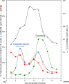
Quantification of a sucrose gradient showing the distribution of Syn5 precursor species. The percent totals of the individual species in fractions 5 to 15 were calculated and plotted against the fractions. Proteins are identified as indicated on their respective axes.

Similar articles

Cited by

References

    1. Shan J, Jia Y, Clokie MR, Mann NH. 2008. Infection by the “photosynthetic” phage S-PM2 induces increased synthesis of phycoerythrin in Synechococcus sp. WH7803. FEMS Microbiol. Lett. 283:154–161. 10.1111/j.1574-6968.2008.01148.x - DOI - PubMed
    1. Lindell D, Jaffe JD, Johnson ZI, Church GM, Chisholm SW. 2005. Photosynthesis genes in marine viruses yield proteins during host infection. Nature 438:86–89. 10.1038/nature04111 - DOI - PubMed
    1. Huiskonen JT, Kivela HM, Bamford DH, Butcher SJ. 2004. The PM2 virion has a novel organization with an internal membrane and pentameric receptor binding spikes. Nat. Struct. Mol. Biol. 11:850–856. 10.1038/nsmb807 - DOI - PubMed
    1. Huiskonen JT, Manole V, Butcher SJ. 2007. Tale of two spikes in bacteriophage PRD1. Proc. Natl. Acad. Sci. U. S. A. 104:6666–6671. 10.1073/pnas.0608625104 - DOI - PMC - PubMed
    1. Waterbury JB, Valois FW. 1993. Resistance to co-occurring phages enables marine Synechococcus communities to coexist with cyanophages abundant in seawater. Appl. Environ. Microbiol. 59:3393–3399 - PMC - PubMed

Publication types

MeSH terms

Substances

LinkOut - more resources