. 2014 Feb;88(4):2071-82.
doi: 10.1128/JVI.02279-13. Epub 2013 Dec 4.

De novo prion aggregates trigger autophagy in skeletal muscle

Affiliations

De novo prion aggregates trigger autophagy in skeletal muscle

Shivanjali Joshi-Barr et al. J Virol. 2014 Feb.

Abstract

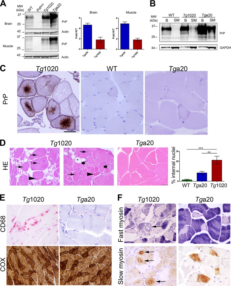
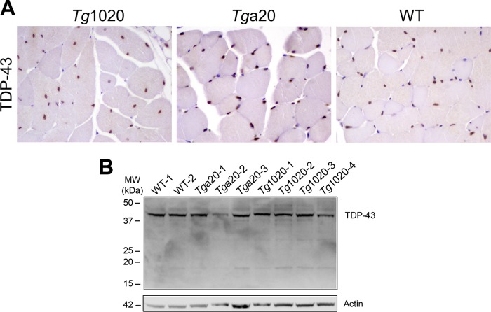
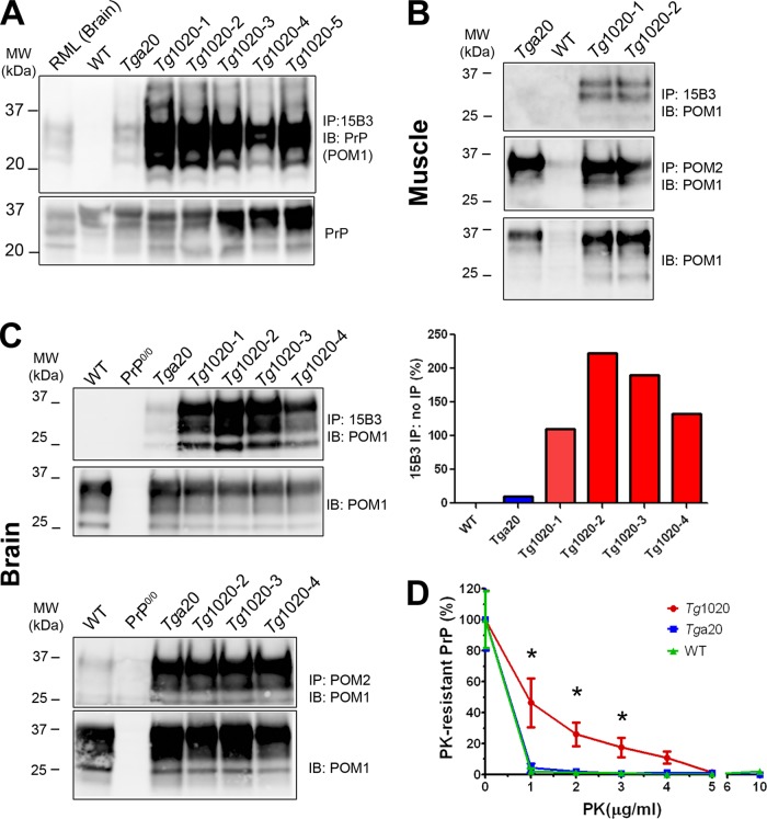
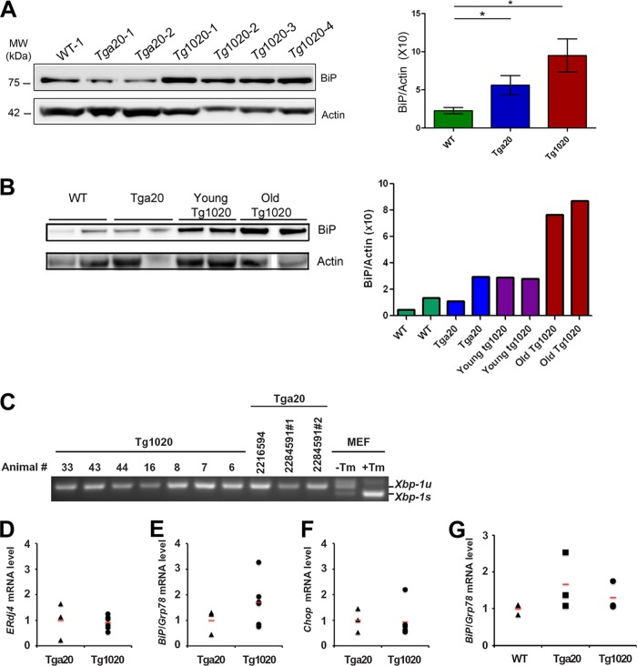
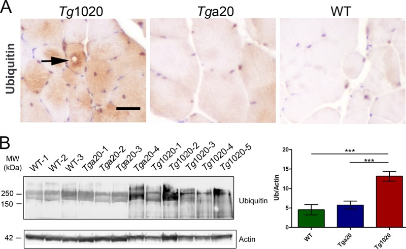
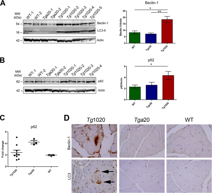
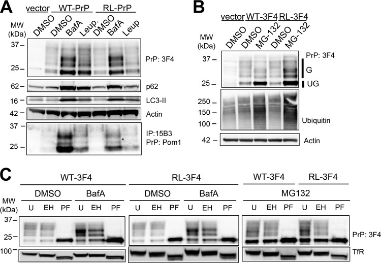
In certain sporadic, familial, and infectious prion diseases, the prion protein misfolds and aggregates in skeletal muscle in addition to the brain and spinal cord. In myocytes, prion aggregates accumulate intracellularly, yet little is known about clearance pathways. Here we investigated the clearance of prion aggregates in muscle of transgenic mice that develop prion disease de novo. In addition to neurodegeneration, aged mice developed a degenerative myopathy, with scattered myocytes containing ubiquitinated, intracellular prion inclusions that were adjacent to myocytes lacking inclusions. Myocytes also showed elevated levels of the endoplasmic reticulum chaperone Grp78/BiP, suggestive of impaired protein degradation and endoplasmic reticulum stress. Additionally, autophagy was induced, as indicated by increased levels of beclin-1 and LC3-II. In C2C12 myoblasts, inhibition of autophagosome maturation or lysosomal degradation led to enhanced prion aggregation, consistent with a role for autophagy in prion aggregate clearance. Taken together, these findings suggest that the induction of autophagy may be a central strategy for prion aggregate clearance in myocytes. IMPORTANCE In prion diseases, the prion protein misfolds and aggregates in the central nervous system and sometimes in other organs, including muscle, yet the cellular pathways of prion aggregate clearance are unclear. Here we investigated the clearance of prion aggregates in the muscle of a transgenic mouse model that develops profound muscle degeneration. We found that endoplasmic reticulum stress pathways were activated and that autophagy was induced. Blocking of autophagic degradation in cell culture models led to an accumulation of aggregated prion protein. Collectively, these findings suggest that autophagy has an instrumental role in prion protein clearance.


Figures

FIG 1
FIG 1
Aged tg1020 mice develop a degenerative myopathy and accumulate central intracellular PrP aggregates in myocytes. (A). Western blot for PrP using equal amounts of protein from each mouse line. (B) Western blot of brain (B) and skeletal muscle (SM) shows higher PrP expression in the brain than in skeletal muscle, even with three times more protein loaded for the muscle samples (note higher GAPDH levels in muscles). POM1 was used for PrP detection. (C) Immunohistochemistry of skeletal muscle revealed coarse, dense perinuclear intracellular PrP inclusions in scattered myocytes of tg1020 mice but not in WT or tga20 mice. (D) Representative images of muscle show central nuclei (arrows), split fibers (arrowheads), endomysial fibrosis (broad arrows), and adipocyte replacement of myocytes (star) in tg1020 mice. HE, hematoxylin and eosin. Asterisks in the bar graph on the right indicate significant differences in the percentages of internal nuclei among the mice by Student's t test (**, P < 0.01; ***, P < 0.001). (E) Focal regions of macrophage infiltration (CD68) were occasionally observed in tg1020 muscle but not in tga20 muscle. A cytochrome oxidase stain (COX) reveals no clear difference in mitochondrial number or pattern between tg1020 and tga20 muscle. (F) Slow and fast myosin heavy chain immunostains show that both type I and type II fibers are degenerating (arrows) in tg1020 skeletal muscle.
FIG 2
FIG 2
TDP-43 expression and localization in WT, tg1020, and tga20 skeletal muscle. (A) Immunostains for TDP-43 in skeletal muscle tissue show that TDP-43 is restricted to nuclei. (B) Western blotting of skeletal muscle using an anti-TDP-43 antibody shows no marked differences in TDP-43 levels among the mice.
FIG 3
FIG 3
PrP aggregates accumulate in tg1020 muscle. (A) (Top) Immunoprecipitation using the PrP conformation-specific antibody 15B3 reveals abundant PrP aggregates in tg1020 skeletal muscle. (Bottom) The protein loaded for the IP contained similar amounts of PrP. Note that 4-fold-larger amounts of the WT sample were loaded. IB, immunoblot; POM1, anti-PrP antibody. (B and C) PrP in muscle and brain, respectively. Antibodies 15B3 and POM2 were used for nondenaturing and denaturing IP, respectively, of the insoluble PrP fraction obtained after ultracentrifugation. POM1 was used for detection by Western blotting. Lower panels show the PrP levels for each sample used for IP. (D) PrP in tg1020 muscle is more PK resistant than PrP in the muscle of tga20 or WT mice as analyzed by PrP ELISA (graphed values are normalized). Asterisks indicate significant differences (*, P ≤ 0.05) by Student's t test.
FIG 4
FIG 4
Induction of the unfolded protein response and an analysis of select ER stress markers in tg1020 skeletal muscle. (A) Western blot analysis using an antibody for UPR marker Grp78/BiP shows an approximately 4-fold-higher level of Grp78/BiP in tg1020 muscle than in WT muscle. Asterisks indicate significant differences (*, P < 0.05) by Student's t test. (B) Young tg1020 mice had slightly higher levels of BiP than WT mice. Note: a tg1020 sample was removed from the blot due to very low actin levels. (C) Semiquantitative real-time PCR reveals no Xbp-1 mRNA splicing in tg1020 skeletal muscle compared to tga20 controls. Xbp-1u and Xbp-1s indicate the unspliced and spliced forms of Xbp-1 mRNA, respectively. Mouse embryonic fibroblasts (MEF) treated with tunicamycin (Tm) showed strong Xbp-1 mRNA splicing and served as a positive control. (D) Quantitative PCR analysis of mRNA levels of ERdj4, a downstream transcriptional target of XBP-1, showed no difference between tga20 and tg1020 animals. (E and F) Quantitative PCR analysis of BiP/Grp78 and Chop mRNA levels, respectively, revealed higher levels of BiP/Grp78 in the muscles of some tg1020 mice than in tga20 mice; however, the differences were not statistically significant. (G) Quantitative PCR analysis revealed no significant difference in BiP/Grp78 mRNA levels between the muscles of young tg1020 mice and those of young WT or tga20 mice. In panels D through F, 3 tga20 and 6 tg1020 animals were analyzed for quantitative PCR measurements, and the values for each animal were plotted individually.
FIG 5
FIG 5
Accumulation of ubiquitinated proteins in skeletal muscle of tg1020 mice (A) Immunohistochemistry shows ubiquitinated inclusions (arrow) in tg1020 muscle. (B) Assessment of total ubiquitinated protein by Western blotting shows an approximately 3-fold-higher level of ubiquitinated proteins in tg1020 muscle than in WT or Tga20 muscle. Asterisks indicate significant differences (***, P < 0.001) by Student's t test.
FIG 6
FIG 6
Increased expression of autophagy-related proteins in tg1020 muscle. (A) Beclin-1 and LC3-II levels were higher in tg1020 muscle than in WT or tga20 muscle. Asterisks indicate significant differences (*, P < 0.05; **, P < 0.01) by Student's t test. (B) Muscle homogenates also showed increased levels of p62 in tg1020 muscle. (C) Quantitative real-time PCR analysis of mRNA in muscles revealed that p62 mRNA levels in tg1020 mice do not differ from those in WT or tga20 mice. (D) Beclin-1 immunostains revealed occasional large central aggregates in tg1020 myocytes, whereas LC3 showed punctate peripheral staining (arrows).
FIG 7
FIG 7
Inhibition of autophagy and proteasomal pathways leads to the accumulation of distinct PrP isoforms in C2C12 cells. (A) Lysates from C2C12 cells transfected either with an empty vector or with a vector encoding WT-PrP or RL-PrP show elevated levels of both unglycosylated and glycosylated PrP upon BafA or leupeptin treatment. Increased p62 and LC3-II levels show the efficacy of the BafA and leupeptin treatments in modulating autophagy. Immunoprecipitation with the PrP conformational antibody 15B3 shows that PrP aggregates accumulate in cells expressing WT-PrP and RL-PrP only upon autophagic inhibition using BafA or leupeptin. (B) Lysates from C2C12 cells show elevated levels of unglycosylated WT-PrP and RL-PrP upon MG132 treatment. Glycosylated and unglycosylated PrP signals are labeled G and UG, respectively. An increase in the level of total ubiquitinated proteins demonstrates the efficacy of MG132 treatment. Immunoblotting for actin shows that equal amounts of protein were loaded. (C) C2C12 cells were transfected with WT-PrP or RL-PrP and were treated with DMSO, BafA, or MG132. Lysates either were left untreated (U) or were treated with endo H (EH) or PNGase F (PF). Glycosylated WT-PrP and RL-PrP that accumulated upon BafA treatment were endo H resistant, yet upon MG132 treatment, they were endo H sensitive. Endo H and PNGase activities were demonstrated by shifts in the molecular weight of the transferrin receptor (TfR) corresponding to the losses of 1 and 2 glycans, respectively, compared to TfR in the untreated samples.

Similar articles

Cited by

References

    1. Prusiner SB. 1982. Novel proteinaceous infectious particles cause scrapie. Science 216:136–144. 10.1126/science.6801762 - DOI - PubMed
    1. Prusiner SB. 1984. Prions: novel infectious pathogens. Adv. Virus Res. 29:1–56. 10.1016/S0065-3527(08)60404-2 - DOI - PubMed
    1. Wadsworth JD, Collinge J. 2011. Molecular pathology of human prion disease. Acta Neuropathol. 121:69–77. 10.1007/s00401-010-0735-5 - DOI - PMC - PubMed
    1. Aguzzi A, Falsig J. 2012. Prion propagation, toxicity and degradation. Nat. Neurosci. 15:936–939. 10.1038/nn.3120 - DOI - PubMed
    1. Deleault NR, Harris BT, Rees JR, Supattapone S. 2007. Formation of native prions from minimal components in vitro. Proc. Natl. Acad. Sci. U. S. A. 104:9741–9746. 10.1073/pnas.0702662104 - DOI - PMC - PubMed

Publication types