. 2014 Feb;88(4):2056-70.
doi: 10.1128/JVI.03051-13. Epub 2013 Dec 4.

Internal ribosome entry site-based attenuation of a flavivirus candidate vaccine and evaluation of the effect of beta interferon coexpression on vaccine properties

Affiliations

Internal ribosome entry site-based attenuation of a flavivirus candidate vaccine and evaluation of the effect of beta interferon coexpression on vaccine properties

Michael Frese et al. J Virol. 2014 Feb.

Abstract

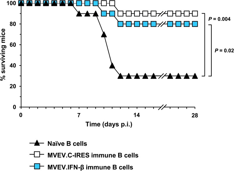
Infectious clone technologies allow the rational design of live attenuated viral vaccines with the possibility of vaccine-driven coexpression of immunomodulatory molecules for additional vaccine safety and efficacy. The latter could lead to novel strategies for vaccine protection against infectious diseases where traditional approaches have failed. Here we show for the flavivirus Murray Valley encephalitis virus (MVEV) that incorporation of the internal ribosome entry site (IRES) of Encephalomyocarditis virus between the capsid and prM genes strongly attenuated virulence and that the resulting bicistronic virus was both genetically stable and potently immunogenic. Furthermore, the novel bicistronic genome organization facilitated the generation of a recombinant virus carrying an beta interferon (IFN-β) gene. Given the importance of IFNs in limiting virus dissemination and in efficient induction of memory B and T cell antiviral immunity, we hypothesized that coexpression of the cytokine with the live vaccine might further increase virulence attenuation without loss of immunogenicity. We found that bicistronic mouse IFN-β coexpressing MVEV yielded high virus and IFN titers in cultured cells that do not respond to the coexpressed IFN. However, in IFN response-sufficient cell cultures and mice, the virus produced a self-limiting infection. Nevertheless, the attenuated virus triggered robust innate and adaptive immune responses evidenced by the induced expression of Mx proteins (used as a sensitive biomarker for measuring the type I IFN response) and the generation of neutralizing antibodies, respectively. IMPORTANCE The family Flaviviridae includes a number of important human pathogens, such as Dengue virus, Yellow fever virus, Japanese encephalitis virus, West Nile virus, and Hepatitis C virus. Flaviviruses infect large numbers of individuals on all continents. For example, as many as 100 million people are infected annually with Dengue virus, and 150 million people suffer a chronic infection with Hepatitis C virus. However, protective vaccines against dengue and hepatitis C are still missing, and improved vaccines against other flaviviral diseases are needed. The present study investigated the effects of a redesigned flaviviral genome and the coexpression of an antiviral protein (interferon) on virus replication, pathogenicity, and immunogenicity. Our findings may aid in the rational design of a new class of well-tolerated and safe vaccines.


Figures

FIG 1
FIG 1
Schematic representation of the wild-type MVEV genome and two genetically engineered, bicistronic MVEV-based constructs. (A) MVEV.wt has a single, long open reading frame encoding all structural (C, prM, and E) and nonstructural (NS1 to NS5) proteins and flanked by untranslated regions (5′ and 3′ UTR). (B) For construction of MVEV.C-IRES, an opal stop codon (filled triangle) was introduced at the C terminus of C protein, followed by an EMCV IRES, which drives translation of all other viral proteins, starting at the signal peptide of prM (black box). (C) Construct MVEV.IFN-β has the mouse IFN-β gene (blue box) inserted downstream of the prM signal peptide, which directs the nascent IFN protein into the lumen of the ER; the IFN-β gene is flanked at the 3′ end by an opal stop codon (filled triangle), followed by EMCV IRES sequence (yellow arrow). The amino acid sequence (single-letter code) and corresponding nucleotide sequence of the prM signal peptide are shown at the bottom (60). Note that synonymous nucleotide changes were introduced into the prM signal peptide upstream of the IRES in construct MVEV.IFN-β to prevent homologous recombination and deletion of the foreign sequences from the viral genome (see sequence alignment; the top and bottom nucleotide sequences encode the wild-type and the altered prM signal peptide, respectively; the resulting amino acid sequence is in uppercase letters, and dots represent identical residues).
FIG 2
FIG 2
Recombinant IFN expression from a bicistronic flavivirus. (A) Vero cells grown in 6-well culture trays (5 × 10⁵ cells/well) were infected with MVEV.IFN-β (IFN-β) or the control virus MVEV.C-IRES (C.IRES) at a multiplicity of 5 PFU per cell. At 24 h p.i., the entire culture supernatant (1 ml) was harvested and replaced with 1 ml of fresh medium, a second harvest was taken at 48 h p.i., and the IFN-β protein concentration in both samples was determined by ELISA. Means from 2 samples ± standard deviations (SD) are presented. (B) Wild-type B6 MEFs in 6-well culture trays (5 × 10⁵ cells/well) were treated for 16 h with 1.5 ml of 102- or 103-fold dilutions of UV-inactivated culture fluid that was harvested 48 h p.i. from Vero cells infected with MVEV.IFN-β or MVEV.C-IRES. After this pretreatment, MEFs were infected with Semliki Forest virus (multiplicity of infection of ∼1), and 24 h later, virus yields were measured by plaque titration on Vero cells. Means from 3 samples ± SD are presented.
FIG 3
FIG 3
Growth of MVEV.wt and two bicistronic MVEV-based viruses in cell culture. (A) Plaque morphologies for MVEV.wt, MVEVC.IRES, and MVEV.IFN-β on Vero cell monolayers. (B, C, and D) Growth kinetics in Vero cells, IFN-sufficient (wt), and defective (IFNAR−/−) MEFs, respectively. Cells were infected at a multiplicity of infection of 0.1 (Vero), 1 (wt MEFs), or 0.2 (IFNAR−/− MEFs); in a double-infection experiment (blue diamonds), MEFs were simultaneously infected with MVEV.wt and MVEV.IFN-β (with a multiplicity of infection of 1 for each virus). Virus titers in the cell culture fluid were determined by plaque titration on Vero cells. The dotted lines denote the detection limit of virus yield by plaque assay. The mean titers ± standard errors of the means (SEM) from two independent experiments are shown.
FIG 4
FIG 4
Genome stability of MVEV.IFN-β after serial passaging. (A) Schematic diagram of the experimental setup to measure the genetic stability of MVEV.IFN-β. (B) Electrophoresis profiles of RT-PCR products of MVEV.wt (lane 1) and MVEV.IFN-β after cell passages 2, 5, 7, 8, 9, and 10 (lanes 2 to 7, respectively). Positions of marker bands and expected product sizes are shown on the left and right, respectively.
FIG 5
FIG 5
Virus attenuation in immunodeficient mice. (A and B) Groups of 6- to 7-week-old NOD-scid mice or 6- to 8-week-old IFNAR−/− mice were infected with 103 to 105 PFU i.v. (scid mice) or 102 to 105 PFU i.p. (IFNAR−/− mice) of MVEV.wt, MVEV.C.IRES, or MVEV.IFN-β. Mice were monitored daily for a period of 28 days and morbidity and mortality were recorded. (C) At 2 days p.i., sera were collected from MVEV.C-IRES (n = 8)- and MVEV.IFN-β (n = 9)-infected IFNAR−/− mice and viremia titers determined by plaque assay on Vero cells. Each symbol represents an individual mouse, and mean titers are indicated by a horizontal red line. The dotted line represents the detection limit of the plaque assay.
FIG 6
FIG 6
Virus RNA and innate immune mRNA quantification in the brain. BALB.A2G-Mx1 mice (3 weeks old) were intracranially inoculated with 104 PFU of MVEV.wt, MVEV.C-IRES, or MVEV.IFN-β. Groups of 2 or 3 mice were killed at the time points indicated, and total RNA was isolated from a quarter of the brain. Quantitative RT-PCR was performed for MVEV RNA (A), IFN-β mRNA (B), and Mx1 mRNA (C). Bars represent mean values obtained from three independent quantitative PCRs (A) or are representative values chosen from two independent quantitative PCRs (B and C). GE, genome equivalent.
FIG 7
FIG 7
Virus and Mx1 protein accumulation in the brains of MVEV-infected mice. Adult BALB.A2G-Mx1 mice were intracranially inoculated with 104 PFU of MVEV.wt (mouse B1-4) (A), diluent alone (mock, mouse A1-1) (B), MVEV.C-IRES (mouse C1-4) (C), and MVEV.IFN-β (mouse D1-4) (D). Mice were killed at 4 days p.i., 4-μm sagittal brain sections were prepared, and neighboring sections were immunostained for the virus protein NS1 (anti-NS1) or Mx1 (anti-Mx1). Sections were then counterstained using either hematoxylin (together with anti-NS1) or eosin (with anti-Mx1). Insets show the typical cytoplasmic and nuclear staining patterns of NS1 and Mx1, respectively. Arrows point to a number of NS1-positive cells in the olfactory bulb of mouse C1-4. Bars, 200 μm.
FIG 8
FIG 8
Protection against lethal challenge with MVEV. B6 donor mice (10 mice/group) were immunized (i.m.) with two doses of 105 PFU of either MVEV.C-IRES or MVEV.IFN-β, delivered 3 weeks apart (Table 3), and B cells were isolated at 9 weeks after completion of the vaccination schedule and adoptively transferred into 4-week-old recipients (n = 10/group). Additionally, B cells from naive mice were transferred into control mice (n = 10). One day after the transfer, mice were challenged with 105 PFU of MVEV.wt by footpad injection and monitored twice daily for morbidity and mortality for 28 days.

Similar articles

Cited by

References

    1. Coleman JR, Papamichail D, Skiena S, Futcher B, Wimmer E, Mueller S. 2008. Virus attenuation by genome-scale changes in codon pair bias. Science 320:1784–1787. 10.1126/science.1155761 - DOI - PMC - PubMed
    1. Yang C, Skiena S, Futcher B, Mueller S, Wimmer E. 2013. Deliberate reduction of hemagglutinin and neuraminidase expression of influenza virus leads to an ultraprotective live vaccine in mice. Proc. Natl. Acad. Sci. U. S. A. 110:9481–9486. 10.1073/pnas.1307473110 - DOI - PMC - PubMed
    1. Plante K, Wang E, Partidos CD, Weger J, Gorchakov R, Tsetsarkin K, Borland EM, Powers AM, Seymour R, Stinchcomb DT, Osorio JE, Frolov I, Weaver SC. 2011. Novel chikungunya vaccine candidate with an IRES-based attenuation and host range alteration mechanism. PLoS Pathog. 7:e1002142. 10.1371/journal.ppat.1002142 - DOI - PMC - PubMed
    1. Guerbois M, Volkova E, Forrester NL, Rossi SL, Frolov I, Weaver SC. 2013. IRES-driven expression of the capsid protein of the Venezuelan equine encephalitis virus TC-83 vaccine strain increases its attenuation and safety. PLoS Negl. Trop. Dis. 7:e2197. 10.1371/journal.pntd.0002197 - DOI - PMC - PubMed
    1. Rossi SL, Guerbois M, Gorchakov R, Plante KS, Forrester NL, Weaver SC. 2013. IRES-based Venezuelan equine encephalitis vaccine candidate elicits protective immunity in mice. Virology 437:81–88. 10.1016/j.virol.2012.11.013 - DOI - PMC - PubMed

Publication types

MeSH terms