. 2014 Feb;160(Pt 2):446-457.
doi: 10.1099/mic.0.072835-0. Epub 2013 Dec 4.

The FupA/B protein uniquely facilitates transport of ferrous iron and siderophore-associated ferric iron across the outer membrane of Francisella tularensis live vaccine strain

Affiliations

The FupA/B protein uniquely facilitates transport of ferrous iron and siderophore-associated ferric iron across the outer membrane of Francisella tularensis live vaccine strain

Girija Ramakrishnan et al. Microbiology (Reading). 2014 Feb.

Abstract

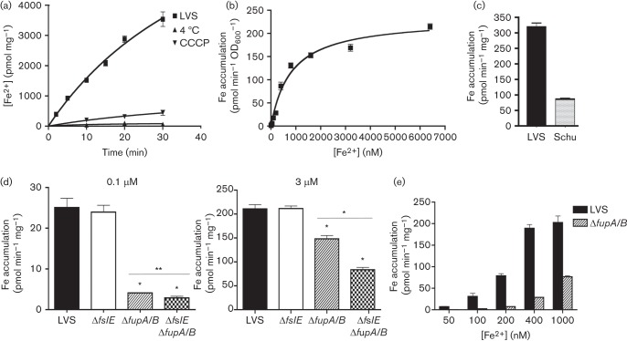
Francisella tularensis is a highly infectious Gram-negative pathogen that replicates intracellularly within the mammalian host. One of the factors associated with virulence of F. tularensis is the protein FupA that mediates high-affinity transport of ferrous iron across the outer membrane. Together with its paralogue FslE, a siderophore-ferric iron transporter, FupA supports survival of the pathogen in the host by providing access to the essential nutrient iron. The FupA orthologue in the attenuated live vaccine strain (LVS) is encoded by the hybrid gene fupA/B, the product of an intergenic recombination event that significantly contributes to attenuation of the strain. We used (55)Fe transport assays with mutant strains complemented with the different paralogues to show that the FupA/B protein of LVS retains the capacity for high-affinity transport of ferrous iron, albeit less efficiently than FupA of virulent strain Schu S4. (55)Fe transport assays using purified siderophore and siderophore-dependent growth assays on iron-limiting agar confirmed previous findings that FupA/B also contributes to siderophore-mediated ferric iron uptake. These assays further demonstrated that the LVS FslE protein is a weaker siderophore-ferric iron transporter than the orthologue from Schu S4, and may be a result of the sequence variation between the two proteins. Our results indicate that iron-uptake mechanisms in LVS differ from those in Schu S4 and that functional differences in the outer membrane iron transporters have distinct effects on growth under iron limitation.


Figures

Fig. 1.
Fig. 1.
Ferrous iron uptake in LVS and mutants. (a) Kinetics of 55Fe2+ transport by LVS. LVS bacteria were grown in iron-limiting che-CDM for 16 h and then diluted and further grown under iron limitation for 22 h. The bacteria were washed and incubated with 7.4 µM 55Fe2+ at 37 °C and incorporation of 55Fe2+ over time was determined by scintillation counting. Transport reactions were carried out in parallel at 4 °C and with bacteria that had been pretreated with carbonyl cyanide m-chlorophenyl hydrazone. (b) Rate of 55Fe2+ transport by LVS plotted as a function of ferrous iron concentration in the uptake reaction. (c) Rate of 55Fe2+ transport by LVS and Schu S4 at 3 micromol ferrous iron. (d) Rates of high-affinity (0.1 µM) and low-affinity (3 µM) ferrous iron transport in LVS and the ΔfslE and ΔfupA/B mutants after growth in iron-limiting media for 20 h. (e) Comparison of ferrous iron transport rates of LVS and ΔfupA/B mutant over a range of iron concentrations. 55Fe accumulation was normalized to protein content or to cell density (OD600). Values are plotted as means with se. Significance was calculated relative to LVS values: *P<0.001, **P<0.02.
Fig. 2.
Fig. 2.
Ferrous iron uptake in fupA/B and fupA complemented strains. Rates of high-affinity (0.1 µM) and low-affinity (3 µM) ferrous iron transport in (a) LVS ΔfslE ΔfupA/B and (b) Schu ΔfslE ΔfupA complemented in cis with fupA/B, fupA and fupB as indicated. Vector indicates control strains in which the vector plasmid alone was integrated in the chromosome. Values are plotted as means with se; *P<0.01; **P<0.001.
Fig. 3.
Fig. 3.
Expression of fupA and fupA/B in complemented strains. Lysates of bacteria grown in iron-replete (H) or iron-limiting (L) media were analysed by SDS-PAGE and Western blotting with antibodies to FupA (FupA/B) and control FipB as indicated. The arrowhead indicates the FupA band. Vector indicates strains carrying the control vector plasmid. (a) Whole-cell lysates of LVS ΔfslE ΔfupA/B complemented with different genes as indicated were tested for expression of FupA (FupA/B). (b) Whole-cell lysates of Schu ΔfslE ΔfupA complemented with fupA and fupA/B mutants were tested for levels of FupA (FupA/B). Corresponding outer membrane proteins (1 µg) were analysed by Western blotting for levels of FupA (FupA/B) and for control protein FipB.
Fig. 4.
Fig. 4.
Growth of LVS ΔfupA/B and LVS ΔfslE ΔfupA/B complements on iron-limiting agar. Tenfold serial dilutions of LVS ΔfupA/B or LVS ΔfslE ΔfupA/B complemented with different genes as indicated were spotted on iron-replete (MHA) or iron-limiting (CDM-Fe) plates. LVS carrying the vector alone was used as control. Growth was recorded after 2 days (MHA) or 4 days (CDM-Fe).
Fig. 5.
Fig. 5.
Growth of Schu ΔfslE ΔfupA complements on iron-limiting agar. Serial dilutions of Schu ΔfslE ΔfupA complemented with endogenous fslE (fslESchu), fupA or fupA/B as indicated were tested for growth on iron-replete (MHA) or on iron-limiting (CDM-Fe) agar as in Fig. 4. Growth of the control Schu S4 bearing the vector alone is shown.
Fig. 6.
Fig. 6.
Growth promotion by cross-feeding of siderophore. (a) Growth promotion of complements by LVS: 3×10⁵ c.f.u. of bacterial strains as indicated were seeded on iron-limiting CDM agar and siderophore-producing LVS bacteria carrying the vector control were spotted in the centre. Growth halo formation around the spot was recorded after 3 days at 37 °C. (b–e) Siderophore dependence of growth halo formation. Bacteria as indicated were seeded on iron-limiting agar and tested for the ability to form growth haloes around spots of siderophore- producing LVS (left) and siderophore-deficient ΔfslA bacteria (right) individually spotted on the same plates. The strains were seeded at titres as follows: (b) 3×10⁵ c.f.u. LVS; (c) 3×10⁶ c.f.u. LVS ΔfslE ΔfupA/B complemented with fupA/B; (d) 3×10⁷ c.f.u. LVS ΔfslE ΔfupA/B complemented with fslELVS; (e) 3×10⁶ c.f.u. LVS ΔfslE ΔfupA/B complemented with fslESchu.
Fig. 7.
Fig. 7.
Expression of fslE in bacterial strains. Whole-cell lysates and outer membrane preparations from bacteria as indicated were analysed by Western blotting for expression of FslE and FupA/B. FipB was used as loading control. (a) Lysates of LVS and ΔfslE and ΔfupA/B mutants and corresponding outer membrane proteins (5 µg) were analysed. (b) Lysates of LVS ΔfslE ΔfupA/B complemented with fslE derived from LVS (fslELVS) or from Schu S4 (fslESchu) and corresponding outer membrane proteins (2 µg) were analysed.
Fig. 8.
Fig. 8.
Siderophore-mediated 55Fe transport. (a) Siderophore-dependent accumulation of 55Fe by LVS. LVS bacteria were grown for 16 h under iron limitation, and diluted and further grown for 24 h in iron-limiting media. The bacteria were washed and incubated with 55Fe in the presence (+sid) or absence (-sid) of siderophore and incorporation of 55Fe over time was determined by scintillation counting. (b) Siderophore-mediated 55Fe transport rates in LVS and ΔfslE, ΔfupA/B or ΔfslE ΔfupA/B mutants grown in iron-limiting media for 20 h. (c) Rates of siderophore-mediated 55Fe transport in LVS ΔfupA/B complemented with different genes as indicated. (d) and (e) Rates of siderophore-mediated 55Fe transport in LVS ΔfslE ΔfupA/B complemented by fupA/B and by fslE orthologues from the LVS and Schu S4 backgrounds. (f) Rates of siderophore-mediated 55Fe transport in Schu ΔfslE ΔfupA/B complemented with different genes as indicated. 55Fe accumulation was normalized to protein content. Values are plotted as means with se. Significance was calculated relative to LVS values in (b) and (c) and relative to vector control in (d), (e) and (f): #P<0.05, *P<0.01, **P<0.002, ***P<0.001; n.s., not significant.

Similar articles

Cited by

References

    1. Bagos P. G., Liakopoulos T. D., Spyropoulos I. C., Hamodrakas S. J. (2004a). PRED-TMBB: a web server for predicting the topology of beta-barrel outer membrane proteins. Nucleic Acids Res 32 (Web Server issue), W400–4. - PMC - PubMed
    1. Bagos P. G., Liakopoulos T. D., Spyropoulos I. C., Hamodrakas S. J. (2004b). A Hidden Markov Model method, capable of predicting and discriminating beta-barrel outer membrane proteins. BMC Bioinformatics 5, 29. 10.1186/1471-2105-5-29 - DOI - PMC - PubMed
    1. Broekhuijsen M., Larsson P., Johansson A., Byström M., Eriksson U., Larsson E., Prior R. G., Sjöstedt A., Titball R. W., Forsman M. (2003). Genome-wide DNA microarray analysis of Francisella tularensis strains demonstrates extensive genetic conservation within the species but identifies regions that are unique to the highly virulent F. tularensis subsp. tularensis. J Clin Microbiol 41, 2924–2931. 10.1128/JCM.41.7.2924-2931.2003 - DOI - PMC - PubMed
    1. Burke D. S. (1977). Immunization against tularemia: analysis of the effectiveness of live Francisella tularensis vaccine in prevention of laboratory-acquired tularemia. J Infect Dis 135, 55–60. 10.1093/infdis/135.1.55 - DOI - PubMed
    1. Chamberlain R. E. (1965). Evaluation of live tularemia vaccine prepared in a chemically defined medium. Appl Microbiol 13, 232–235. - PMC - PubMed

Publication types

MeSH terms

LinkOut - more resources