. 2014 Jan 1;192(1):492-502.
doi: 10.4049/jimmunol.1301812. Epub 2013 Dec 4.

MPYS/STING-mediated TNF-α, not type I IFN, is essential for the mucosal adjuvant activity of (3'-5')-cyclic-di-guanosine-monophosphate in vivo

Affiliations

MPYS/STING-mediated TNF-α, not type I IFN, is essential for the mucosal adjuvant activity of (3'-5')-cyclic-di-guanosine-monophosphate in vivo

Steven M Blaauboer et al. J Immunol. .

Abstract

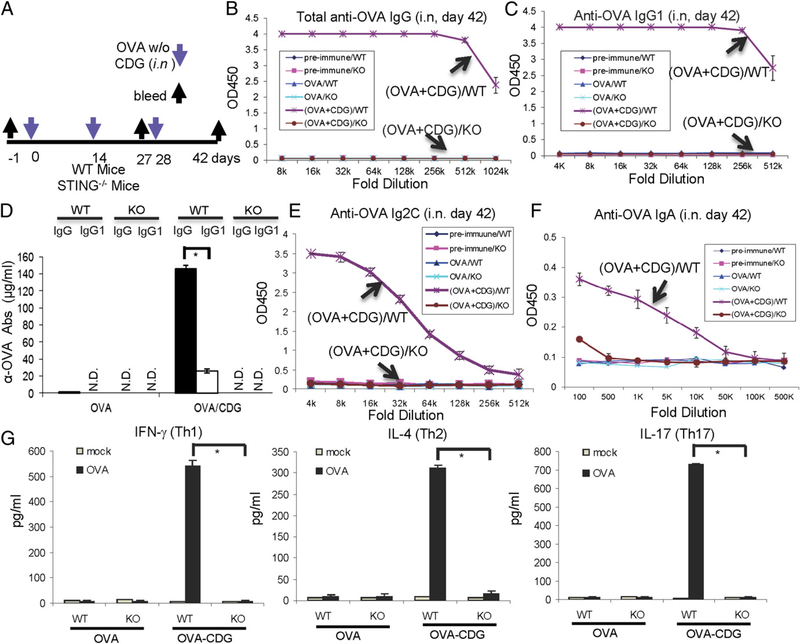
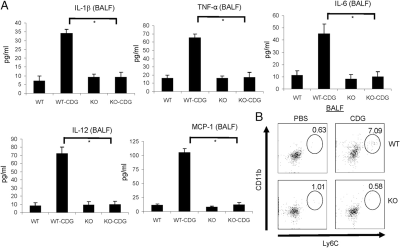
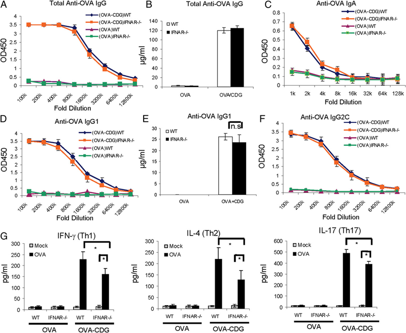
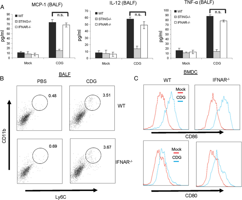
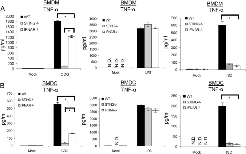
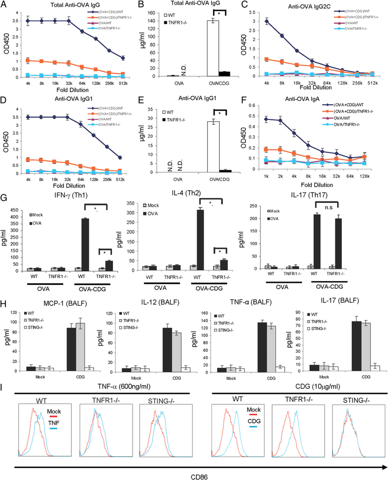
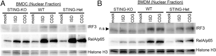
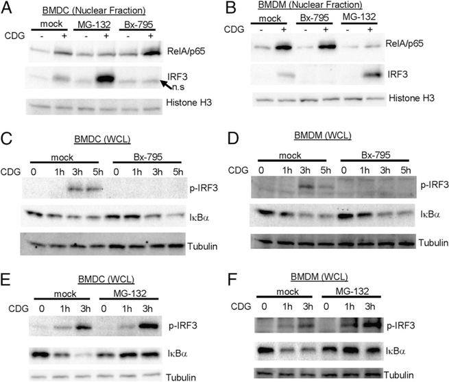
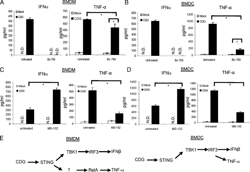
The bacterial second messenger (3'-5')-cyclic-di-guanosine-monophosphate (CDG) is a promising mucosal adjuvant candidate that activates balanced Th1/Th2/Th17 responses. We showed previously that CDG activates stimulator of IFN genes (STING)-dependent IFN-I production in vitro. However, it is unknown whether STING or IFN-I is required for the CDG adjuvant activity in vivo. In this study, we show that STING(-/-) mice (Tmem173(<tm1Camb>)) do not produce Ag-specific Abs or Th1/Th2/Th17 cytokines during CDG/Ag immunization. Intranasal administration of CDG did not induce TNF-α, IL-1β, IL-6, IL-12, or MCP-1 production in STING(-/-) mice. Surprisingly, we found that the cytokine and Ab responses were unaltered in CDG/Ag-immunized IFNAR(-/-) mice. Instead, we found that CDG activates STING-dependent, IFN-I-independent TNF-α production in vivo and in vitro. Furthermore, using a TNFR1(-/-) mouse, we demonstrate that TNF-α signaling is critical for CDG-induced Ag-specific Ab and Th1/Th2 cytokine production. This is distinct from STING-mediated DNA adjuvant activity, which requires IFN-I, but not TNF-α, production. Finally, we found that CDG activates STING-dependent, but IRF3 stimulation-independent, NF-κB signaling. Our results established an essential role for STING-mediated TNF-α production in the mucosal adjuvant activity of CDG in vivo and revealed a novel IFN-I stimulation-independent STING-NF-κB-TNF-α pathway.


Conflict of interest statement

Disclosures

The authors have no financial conflicts of interest.

Figures

FIGURE 1.
FIGURE 1.
STING is essential for the mucosal adjuvant activity of CDG. (A) Outline of the study. Mice were vaccinated i.n. with three doses (14 d apart) of OVA (20 μg) together with CDG (5 μg) or OVA alone (20 μg). Blood and nasal wash samples were collected 14 d after the last immunization (day 42). (B–F) Each group has four mice. Sera or nasal washes from the four mice in the same group were pooled. OVA-specific Abs in the sera and nasal washes were measured by ELISA, as described in Materials and Methods (n = 3). (G) Splenocytes were collected 14 d after the last immunization and were activated in vitro with OVA (100 μg/ml) for 4 d. Supernatants from the same group were pooled together. Th1 (IFN-γ), Th2 (IL-4), and Th17 (IL-17) cytokines were measured by ELISA (n = 3). Data are mean ± SE from three independent experiments. *p, 0.05.
FIGURE 2.
FIGURE 2.
STING is essential for CDG-induced cytokine and chemokine production in vivo. (A) Mice were administered CDG (10 μg) or PBS i.n. After 15 h, cytokine production was measured in BALF by ELISA (n = 3). Data are mean ± SE from three independent experiments. (B) Cells from BALF were analyzed by FACScan with the indicated Abs. Live cells were gated (n = 3). *p < 0.05.
FIGURE 3.
FIGURE 3.
IFN-I signaling is dispensable for the mucosal adjuvant activity of CDG. (A–E) WT and IFNAR−/− mice were vaccinated i.n. as in Fig. 1. Abs in the blood and nasal wash samples were determined as in Fig. 1 (n = 3). (F) Splenocytes from WT and IFNAR−/− mice were collected and activated in vitro with OVA (100 μg/ml) as in Fig. 1. (G) Th1/Th2/Th17 cytokines were determined in supernatants as in Fig. 1 (n = 3). Data are mean ± SE from three independent experiments. *p, 0.05. n.s, Nonsignificant
FIGURE 4.
FIGURE 4.
IFN-I signaling is dispensable for CDG-induced cytokine and chemokine production in vivo. (A) WT, STING−/−, and IFNAR−/− mice were given CDG (10 μg) or PBS i.n. Cytokine production was measured in BALF as in Fig. 2 (n = 3). Data are mean ± SE from three independent experiments. (B) Live cells from BALF were analyzed by FACScan with the indicated Abs as in Fig. 2 (n = 3). (C) BMDCs from WTand IFNAR−/− μiχε were activated by CDG (10 μg/ml) or medium alone (mock) for 18 h. Cells were stained with Abs against CD80 and CD86. Live cells were analyzed by flow cytometry (n = 3). *p, 0.05. n.s., Nonsignificant.
FIGURE 5.
FIGURE 5.
STING differentially regulates CDG- and ISD-induced TNF-α production in vitro. BMDMs (A) or BMDCs (B) from WT, STING−/−, or IFNAR−/− mice were activated by CDG (10 μg/ml), LPS (200 ng/ml), ISD (10 μg/ml), or medium alone (mock) for 6 h. TNF-α production was measured in the supernatant by ELISA (n = 3). Data are mean ± SE from three independent experiments. *p <, 0.05. N.D., Not detected.
FIGURE 6.
FIGURE 6.
TNF-α signaling is critical for the mucosal adjuvant activity of CDG. (A–F) WTand TNFR1−/− mice were vaccinated i.n. as in Fig. 1. Abs in the blood and nasal wash samples were determined as in Fig. 1 (n = 3). (G) Splenocytes from WTand TNFR1−/− mice were collected and activated in vitro with OVA (100 μg/ml) as in Fig. 1. Th1/Th2/Th17 cytokines were determined in supernatants as in Fig. 1 (n = 3). Data are mean ± SE from three independent experiments. (H) WT, STING−/−, and TNFR1−/− mice were treated as in Fig. 2. Cytokine production was measured in BALF as in Fig. 2 (n = 3). Data are mean ± SE from three independent experiments. (I) BMDCs from WT, STING−/−, or TNFR1−/− mice were activated by TNF (600 ng/ml), CDG (10 μg/ml), or medium alone (mock) for 18 h. Cells were stained with Abs against CD86. Live cells were analyzed by flow cytometry (n = 2). *p<,0.05. N.D., Not detected; n.s, nonsignificant.
FIGURE 7.
FIGURE 7.
STING is required for CDG-induced NF-κB activation. BMDCs (A) or BMDMs (B) from C57BL/6, STING2/+, or STING−/− mice were activated by CDG (10 μg/ml) for 4 h. Nuclear fractions were isolated as described in Materials and Methods. Samples were run on SDS-PAGE gel and probed with the indicated Abs (n = 3). n.s, Nonspecific.
FIGURE 8.
FIGURE 8.
Uncoupling STING-mediated NFkB activation and IRF3 activation. BMDCs (A) or BMDMs (B) from C57BL/6 mice were treated with Bx-795 (1 μM), MG-132 (4 μg/ml), or DMSO (mock) for 2 h. Cells were then activated by CDG (10 μg/ml) for 4 h. Nuclear fractions were isolated and processed as in Fig. 7 (n = 3). BMDCs (C) or BMDMs (D) were treated with Bx-795 (1 μM) or DMSO (mock) as in (A) and (B). Cells were then activated by CDG (10 μg/ml) for the indicated time. WCLs were run on SDS-PAGE gel and probed with the indicated Abs (n = 3). BMDCs (E) or BMDMs (F) were treated with MG-132 (4 μg/ml) or DMSO (mock) as in (A) and (B). Cells were then activated by CDG (10 μg/ml) for the indicated time. WCLs were run on SDS-PAGE gel and probed with the indicated Abs (n = 3). n.s, Nonspecific.
FIGURE 9.
FIGURE 9.
Uncoupling STING-mediated TNF-α and IFN-I production. BMDMs (A) or BMDCs (B) from C57BL/6 mice were treated with Bx-795 (1 mM) or DMSO (untreated) for 2 h. Cells were then activated by CDG (10 μg/ml) for 5 h. TNF-α and IFN-α were measured in supernatant (n = 3). Data are mean ± SE from three independent experiments. BMDMs (C) or BMDCs (D) were treated with MG-132 (4 μg) or DMSO (untreated) for 2 h. Cells were then activated by CDG (10 μg/ml) for 5 h. TNF-α and IFN-α were measured in supernatant (n = 3). Data are mean ± SE from three independent experiments. (E) Uncoupling of CDG–STING–TBK1–IRF3–IFN-I with CDG–STING–NF-κB–TNF-α in BMDMs and BMDCs. *p<, 0.05. N.D., Not detected.

References

    1. Karaolis DK, Means TK, Yang D, Takahashi M, Yoshimura T, Muraille E,Philpott D, Schroeder JT, Hyodo M, Hayakawa Y, et al. 2007. Bacterial c-di-GMP is an immunostimulatory molecule. J. Immunol 178: 2171–2181. - PubMed
    1. Ebensen T, Schulze K, Riese P, Link C, Morr M, and Guzma CA´n. 2007. The bacterial second messenger cyclic diGMP exhibits potent adjuvant properties. Vaccine 25: 1464–1469. - PubMed
    1. Madhun AS, Haaheim LR, Nøstbakken JK, Ebensen T, Chichester J,Yusibov V, Guzman CA, and Cox RJ 2011. Intranasal c-di-GMP-adjuvanted plant-derived H5 influenza vaccine induces multifunctional Th1 CD4+ cells and strong mucosal and systemic antibody responses in mice. Vaccine 29: 4973–4982. - PubMed
    1. Zhao L, KuoLee R, Harris G, Tram K, Yan H, and Chen W 2011. c-di-GMPprotects against intranasal Acinetobacter baumannii infection in mice by chemokine induction and enhanced neutrophil recruitment. Int. Immunopharmacol 11: 1378–1383. - PubMed
    1. Hu DL, Narita K, Hyodo M, Hayakawa Y, Nakane A, and Karaolis DK 2009. c-di-GMP as a vaccine adjuvant enhances protection against systemic methicillin-resistant Staphylococcus aureus (MRSA) infection. Vaccine 27: 4867–4873. - PubMed

Publication types

MeSH terms