

Contrast improvement in sub- and ultraharmonic ultrasound contrast imaging by combining several hammerstein models
- PMID: 24307890
- PMCID: PMC3838838
- DOI: 10.1155/2013/270523
Contrast improvement in sub- and ultraharmonic ultrasound contrast imaging by combining several hammerstein models
Abstract
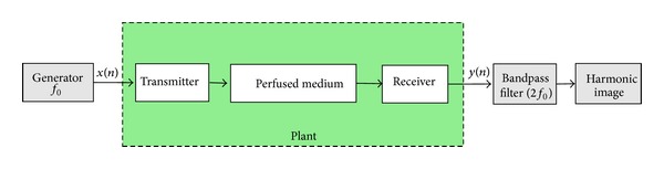
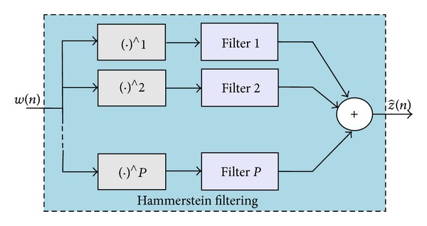
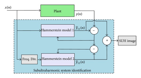
Sub- and ultraharmonic (SUH) ultrasound contrast imaging is an alternative modality to the second harmonic imaging, since, in specific conditions it could produce high quality echographic images. This modality enables the contrast enhancement of echographic images by using SUH present in the contrast agent response but absent from the nonperfused tissue. For a better access to the components generated by the ultrasound contrast agents, nonlinear techniques based on Hammerstein model are preferred. As the major limitation of Hammerstein model is its capacity of modeling harmonic components only, in this work we propose two methods allowing to model SUH. These new methods use several Hammerstein models to identify contrast agent signals having SUH components and to separate these components from harmonic components. The application of the proposed methods for modeling simulated contrast agent signals shows their efficiency in modeling these signals and in separating SUH components. The achieved gain with respect to the standard Hammerstein model was 26.8 dB and 22.8 dB for the two proposed methods, respectively.
Figures


References
-
- Calliada F, Campani R, Bottinelli O, Bozzini A, Sommaruga MG. Ultrasound contrast agents: basic principles. European Journal of Radiology. 1998;27(supplement 2):S157–S160. - PubMed
-
- Forsberg F, Merton DA, Liu JB, Needleman L, Goldberg BB. Clinical applications of ultrasound contrast agents. Ultrasonics. 1998;36(1–5):695–701. - PubMed
-
- Frinking PJA, Bouakaz A, Kirkhorn J, Ten Cate FJ, De Jong N. Ultrasound contrast imaging: current and new potential methods. Ultrasound in Medicine and Biology. 2000;26(6):965–975. - PubMed
-
- Leighton TG. The Acoustic Bubble. London, UK: Academic Press; 1994.
LinkOut - more resources
Full Text Sources
Other Literature Sources
Research Materials
Miscellaneous
