

Clathrin-mediated endocytosis and adaptor proteins
Affiliations
- PMID: 24307937
- PMCID: PMC3848845
Item in Clipboard
Clathrin-mediated endocytosis and adaptor proteins
Acta Naturae.
2013 Jul.
Abstract
Macromolecules gain access to the cytoplasm of eukaryotic cells using one of several ways of which clathrin-dependent endocytosis is the most researched. Although the mechanism of clathrin-mediated endocytosis is well understood in general, novel adaptor proteins that play various roles in ensuring specific regulation of the mentioned process are being discovered all the time. This review provides a detailed account of the mechanism of clathrin-mediated internalization of activated G protein-coupled receptors, as well as a description of the major proteins involved in this process.
Keywords: adaptor proteins; clathrin; endocytosis.
Figures
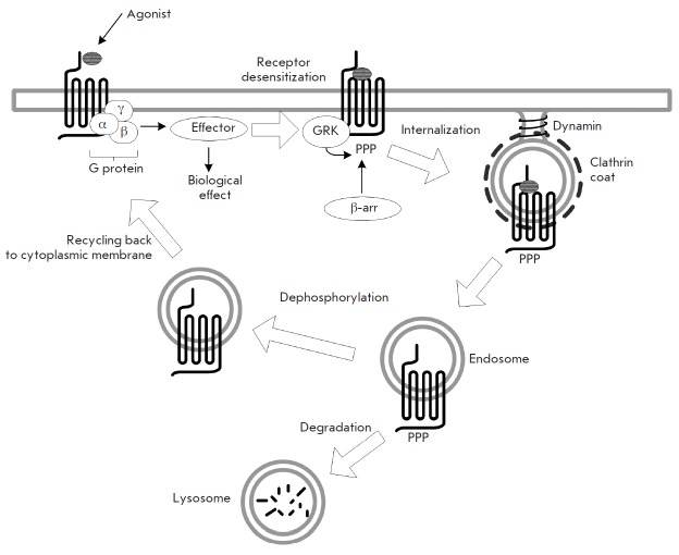
Schematic diagram of clathrin-mediated internalization of a receptor following its activation
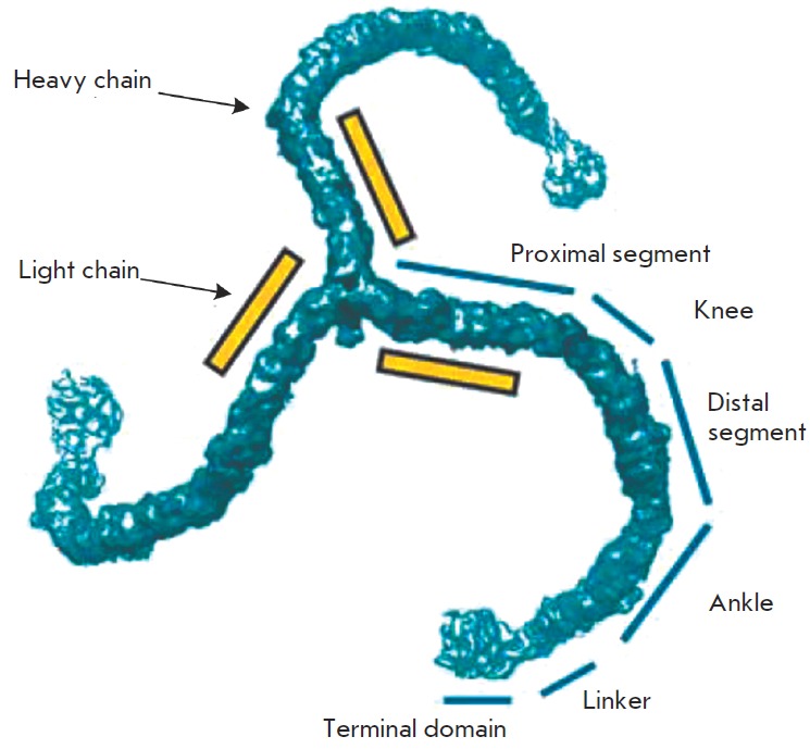
Clathrin molecule (triskelion). Segments of the clathrin heavy chain are
indicated. The terminal domain is the N-terminal domain and the C-terminal
domains are localized in the center of the molecule. The position of the light
chains is shown schematically. Figure adapted from [40]
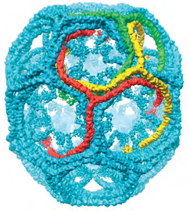
Hexagonal clathrin barrel model (7.9Å resolution). Only the heavy chains of
clathrin are indicated. Figure adapted from [40]
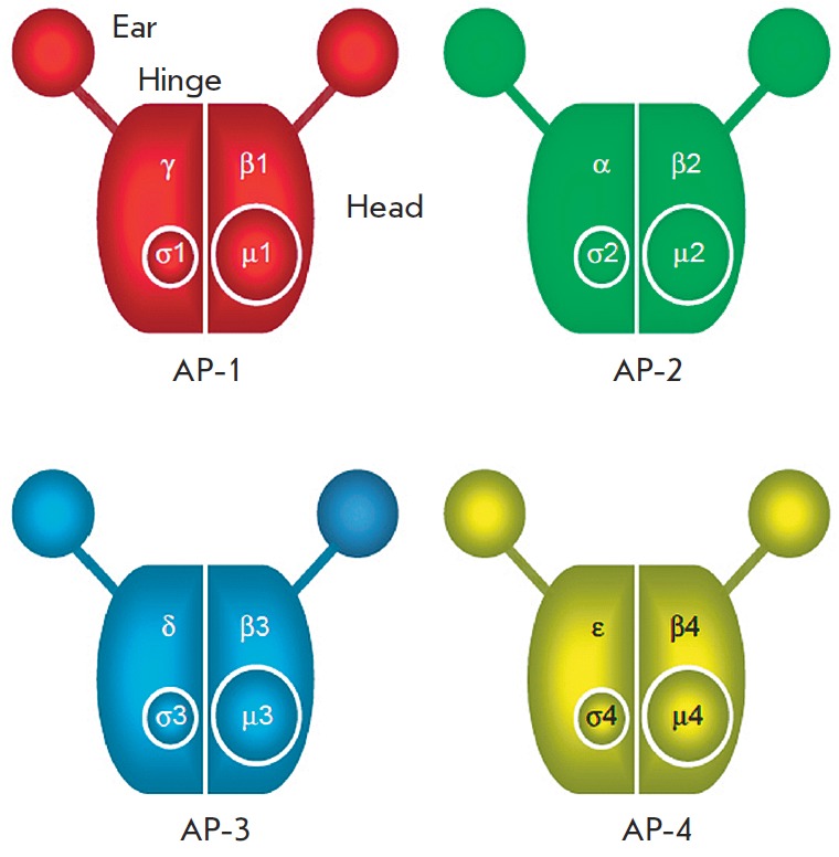
Schematic diagram of the AP complexes. All complexes consist of two large
subunits, one medium subunit, and a small subunit. Figure adapted from [63]
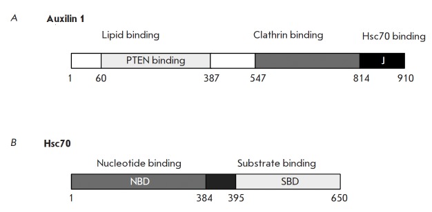
Domain organization of auxilin and Hsc70. Numbers indicate the boundaries of
various domains. Figure adapted from [85]
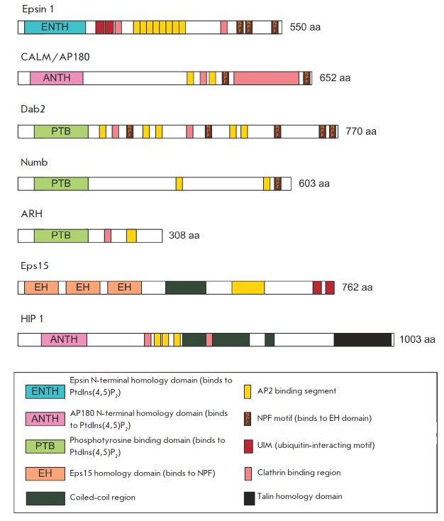
Monomeric clathrin-binding adaptors. Schematic representation of the overall
domain structure. Figure adapted from [70]

Schematic representation of the protein TRIP8b domain organization
References
LinkOut - more resources
Full Text Sources
Other Literature Sources