Review
. 2013 Dec 5:13:75.
doi: 10.1186/1471-2415-13-75.

Alteration of choroidal thickness in a case of carotid cavernous fistula: a case report and a review of the literature

Affiliations
Review

Alteration of choroidal thickness in a case of carotid cavernous fistula: a case report and a review of the literature

Yoichiro Shinohara et al. BMC Ophthalmol. .

Abstract

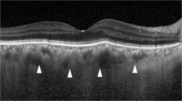
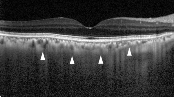
Background: To measure the alterations of the choroidal thickness in Carotid cavernous fistula (CCF) using enhanced depth imaging optical coherence tomography (EDI-OCT).

Case presentation: A 64-year-old woman was referred to us for redness, exophthalmos and visual disturbance in her right eye. She was diagnosed with CCF by magnetic resonance imaging (MRI) and magnetic resonance angiography.Observations; Embolization resulted in improvement of ocular symptoms, and there was a reduction of the subfoveal choroidal thickness in the right eye from 351 μm preoperatively to 142 μm postoperatively in EDI-OCT.

Conclusion: EDI-OCT demonstrated that the choroidal thickness increases occurred due to congestion in a CCF case.


Figures

Figure 1
Figure 1
Left photograph: prior to treatment, the episcleral and conjunctival vessels exhibit tortuosity and dilatation. Right photograph: After treatment, the vessels returned to normal.
Figure 2
Figure 2
Right eye fundus photographs. Left photograph: Prior to treatment, the retinal veins exhibit dilatation and tortuosity due to CCF. Right photograph: After treatment, the venous condition returned to normal.
Figure 3
Figure 3
Enhanced depth imaging optical coherence tomography (EDI-OCT) before and after treatment. Upper image: Prior to treatment, the subfoveal choroidal thickness is 297 μm (arrowheads). Lower image: After treatment, the subfoveal choroidal thickness decreased to 142 μm, which is almost half of the preoperative choroidal thickness (arrowheads).
Figure 4
Figure 4
Preoperative blood flow image obtained by laser speckle flowgraphy Upper fundus image shows the right eye. The fundus color that is primarily seen shows that the choroidal blood flow is cold, which indicates there is a decrease in the choroidal blood speed due to venous congestion. Lower fundus image shows the left eye. In this image, the color is warmer as compared to that observed in the right fundus image.

References

    1. Pillai GS, Ghose S, Singh N, Garodia VK, Puthassery R, Manjunatha NP. Central retinal artery occlusion in dural carotid cavernous fistula. Retina. 2002;22:493–494. doi: 10.1097/00006982-200208000-00015. - DOI - PubMed
    1. Kirsch M, Henkes H, Liebig T, Weber W, Esser J, Golik S, Kuhne D. Endovascular management of dural carotid-cavernous sinus fistulas in 141 patients. Neuroradiology. 2006;48:486–490. doi: 10.1007/s00234-006-0089-9. - DOI - PubMed
    1. Barrow DL, Spector RH, Braun IF, Landman JA, Tindall SC, Tindall GT. Classification and treatment of spontaneous carotid-cavernous sinus fistulas. J Neurosurg. 1985;62:248–256. doi: 10.3171/jns.1985.62.2.0248. - DOI - PubMed
    1. Men S, Ozturk H, Hekimoglu B, Sekerci Z. Traumatic carotid-cavernous fistula treated by combined transarterial and transvenous coil embolization and associated cavernous internal carotid artery dissection treated with stent placement. Case report. J Neurosurg. 2003;99:584–586. doi: 10.3171/jns.2003.99.3.0584. - DOI - PubMed
    1. Hurst RW, Howard RS, Zager E. Carotid cavernous fistula associated with persistent trigeminal artery: endovascular treatment using coil embolization. Skull base surgery. 1998;8:225–228. doi: 10.1055/s-2008-1058188. - DOI - PMC - PubMed

LinkOut - more resources