. 2014 Apr;28(4):446-52.
doi: 10.1089/end.2013.0662. Epub 2014 Jan 8.

Effect of the body wall on lithotripter shock waves

Affiliations

Effect of the body wall on lithotripter shock waves

Guangyan Li et al. J Endourol. 2014 Apr.

Abstract

Purpose: Determine the influence of passage through the body wall on the properties of lithotripter shock waves (SWs) and the characteristics of the acoustic field of an electromagnetic lithotripter.

Methods: Full-thickness ex vivo segments of pig abdominal wall were secured against the acoustic window of a test tank coupled to the lithotripter. A fiber-optic probe hydrophone was used to measure SW pressures, determine shock rise time, and map the acoustic field in the focal plane.

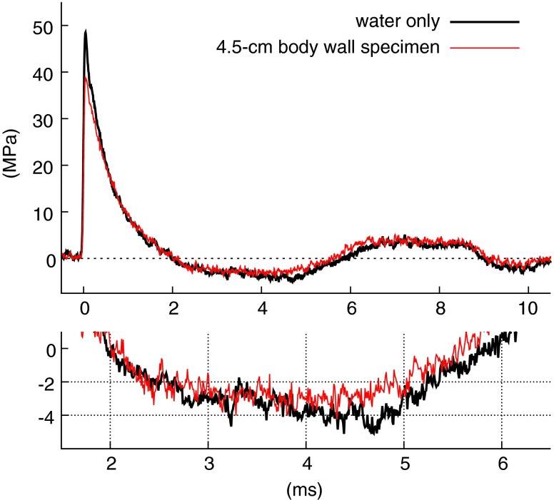
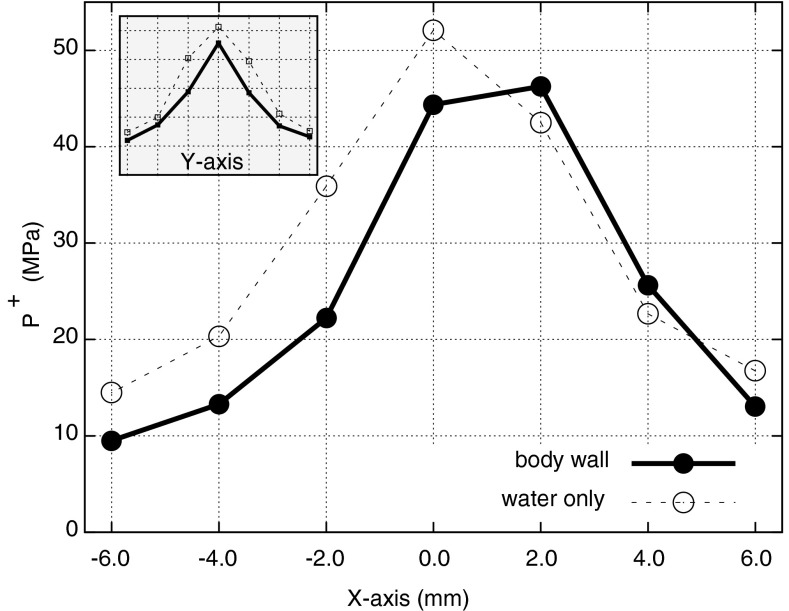
Results: Peak positive pressure on axis was attenuated roughly proportional to tissue thickness-approximately 6% per cm. Irregularities in the tissue path affected the symmetry of SW focusing, shifting the maximum peak positive pressure laterally by as much as ∼2 mm. Within the time resolution of the hydrophone (7-15 ns), shock rise time was unchanged, measuring ∼17-21 ns with and without tissue present. Mapping of the field showed no effect of the body wall on focal width, regardless of thickness of the body wall.

Conclusions: Passage through the body wall has minimal effect on the characteristics of lithotripter SWs. Other than reducing pulse amplitude and having the potential to affect the symmetry of the focused wave, the body wall has little influence on the acoustic field. These findings help to validate laboratory assessment of lithotripter acoustic field and suggest that the properties of SWs in the body are much the same as have been measured in vitro.


Figures

<b>FIG. 1.</b>
FIG. 1.
Ex vivo test system: (a) Measurements were performed in a water-filled test tank using a fiber-optic probe hydrophone (FOPH) moved in the focal plane of the lithotripter by an X-Y-Z positioner. Specimens of pig body wall were held against the Mylar acoustic window of the tank and the treatment head of the lithotripter was coupled to the window using LithoClear® gel. (b) Full-thickness pieces of anterior abdominal wall were used. These consisted of skin (S) and subcutaneous fat (F), the paired rectus abdominis muscles (M) in cross-section view, surrounded by connective tissue fascia (CT) and apposed at the midline, and the peritoneum (P) and its subjacent fascia. Although specimens might be uniform in thickness, they were acoustically non-uniform.
<b>FIG. 2.</b>
FIG. 2.
Representative pressure waveforms for shock waves (SWs) passing through a 4.5 cm-thick ex vivo body wall. The FOPH probe was positioned at the lithotripter focal point during measurement and the two traces shown are the average over 25 SWs. Waveforms with and without the tissue were remarkably similar, showing only a reduction in amplitude.
<b>FIG. 3.</b>
FIG. 3.
Percent attenuation of peak positive pressure (P+) as a function of body wall thickness. Maximum P+ was measured at the lithotripter focal point with nine different body wall specimens (2.5 to 5.5 cm in thickness) at the acoustic window. Linear fit analysis estimates a 6.1% increase in attenuation per centimeter in thickness. Error bars indicate one standard deviation.
<b>FIG. 4.</b>
FIG. 4.
Attenuation of acoustic pressures without an effect on focal width. Mapping of the pressure field along the Y-axis for passage of SWs through a 5.0 cm thick specimen of abdominal wall showed a reduction in both positive pressure (∼41%) and negative pressure (∼25%), but little effect on focal width. Inset: Trace of P+ along the Y-axis for passage of SWs through a thinner (2.5 cm) specimen of body wall showed only a slight attenuation of the pulse and no apparent effect on focal width. The shock-to-shock variation in the 25 consecutive SWs acquired for typical data sets measured <±2.0 MPa without tissue present and <±3.0 MPa with the body wall.
<b>FIG. 5.</b>
FIG. 5.
Disruption in symmetry of the acoustic field by a 3.5 cm thick body wall. Mapping of the field in the X-axis showed lateral displacement of maximum P+ by about 2 mm, while mapping in the Y-axis (inset) showed no such effect. The shock-to-shock variations measured <±2.0 MPa without tissue present and <±3.0 MPa with the tissue.
<b>FIG. 6.</b>
FIG. 6.
Shock rise time for measures close to the tissue. Measures of shock rise time for body wall specimens of different thickness in which the FOPH fiber tip was ∼2–5 cm from the tissue all showed values of ∼17–21 ns (see text). To determine whether this lengthy distance between the FOPH and tissue permitted artifactual healing of the shock front, measures were conducted with the fiber tip in close proximity to the tissue. In this case a specimen of body wall ∼4 cm in thickness was halved and stacked to occupy the space between the acoustic window and the focal point, and the FOPH was advanced to within <0.5 mm of the tissue. Normalized pressure traces for measures with and without tissue show virtually no difference in the shock rise time. For these measures the FOPH was ∼8–9 mm post-focal due to the thickness of the stacked tissue.

Similar articles

Cited by

References

    1. Chaussy C, Schmiedt E, Jocham D, Brendel W, Forssmann B, Walther V. First clinical experience with extracorporeally induced destruction of kidney stones by shock waves. J Urol 1982;127:417–420 - PubMed
    1. Miller NL, Lingeman JE. Treatment of kidney stones: Current lithotripsy devices are proving less effective in some cases. Nat Clin Pract Urol 2006;3:236–237 - PubMed
    1. Ng CF, Lo AKY, Lee KWM, Wong KT, Chung WY, Gohel D. A Prospective, randomized study of the clinical effects of shock wave delivery for unilateral kidney stones: 60 versus 120 shocks per minute. J Urol 2012;188:837–842 - PubMed
    1. Matlaga BR, Lingeman JE. Surgical management of upper urinary tract calculi. In: Wein AJ, Kavoussi LR, Novick AC, Partin AW, Peters CA, eds. Campbell-Walsh Urology. New York, NY: Saunders; 2012, pp 1357–1410
    1. Zhu S, Cocks FH, Preminger GM, Zhong P. The role of stress waves and cavitation in stone comminution in shock wave lithotripsy. Ultrasound Med Biol 2002;28:661–671 - PubMed

Publication types

LinkOut - more resources