. 2013 Dec 5;2(6):e000410.
doi: 10.1161/JAHA.113.000410.

Mechanical dyssynchrony precedes QRS widening in ATP-sensitive K⁺ channel-deficient dilated cardiomyopathy

Affiliations

Mechanical dyssynchrony precedes QRS widening in ATP-sensitive K⁺ channel-deficient dilated cardiomyopathy

Satsuki Yamada et al. J Am Heart Assoc. .

Abstract

Background: Contractile discordance exacerbates cardiac dysfunction, aggravating heart failure outcome. Dissecting the genesis of mechanical dyssynchrony would enable an early diagnosis before advanced disease.

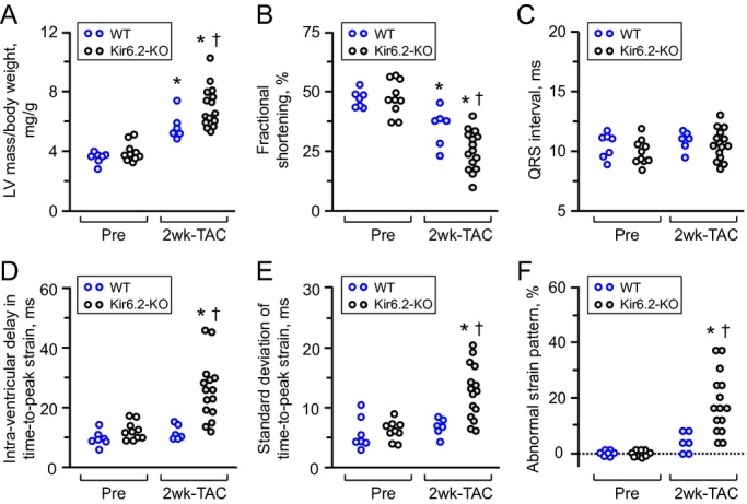
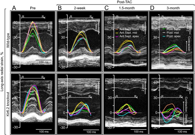
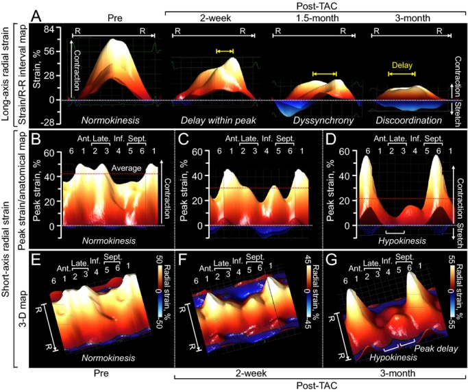
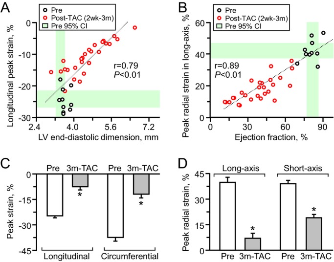
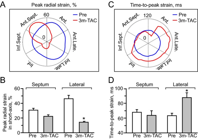
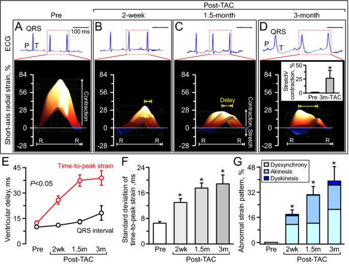
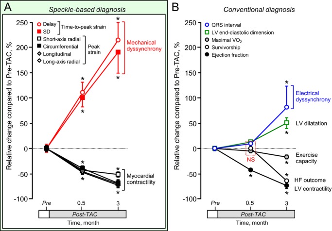
Methods and results: High-resolution speckle-tracking echocardiography was applied in a knockout murine surrogate of adult-onset human cardiomyopathy caused by mutations in cardioprotective ATP-sensitive K(+) (K(ATP)) channels. Preceding the established criteria of cardiac dyssynchrony, multiparametric speckle-based strain resolved nascent erosion of dysfunctional regions within cardiomyopathic ventricles of the K(ATP) channel-null mutant exposed to hemodynamic stress. Not observed in wild-type counterparts, intraventricular disparity in wall motion, validated by the degree, direction, and delay of myocardial speckle patterns, unmasked the disease substrate from asymptomatic to overt heart failure. Mechanical dyssynchrony preceded widening of the QRS complex and exercise intolerance and progressed into global myocardial discoordination and decompensated cardiac pump function, precipitating a low output syndrome.

Conclusions: The present study, with the use of high-resolution imaging, prospectively resolved the origin and extent of intraventricular motion disparity in a K(ATP) channel-knockout model of dilated cardiomyopathy. Mechanical dyssynchrony established as an early marker of cardiomyopathic disease offers novel insight into the pathodynamics of dyssynchronous heart failure.

Keywords: ATP‐sensitive K+ channel; Kir6.2; QRS complex; heart failure; speckle‐tracking.


Figures

Figure 1.
Figure 1.
Hemodynamic stress induced by TAC. Constriction was surgically placed at the level of the transverse aorta (TAC, yellow arrowhead in A). The extent of TAC was determined by reduction in the diameter of the transverse aorta (red in B), which was originally ≈1 mm, and the increase in flow gradient between proximal and distal TAC sites (C). 2‐D/3‐D indicates 2‐/3‐dimensional; As‐Ao, ascending aorta; Des‐Ao, descending aorta; Dist., distal site of TAC; lcca, left common carotid artery; Prox., proximal site of TAC; ria, right innominate artery; TAC, transverse aortic constriction; T‐Ao, transverse aorta; yellow arrows, peak velocity.
Figure 2.
Figure 2.
ATP‐sensitive K+ (KATP) channel knockout aggravates stress‐induced cardiomyopathy associated with mechanical dyssynchrony. Following transverse aortic constriction (TAC), KATP channel‐deficient hearts, due to Kir6.2 subunit knockout (Kir6.2‐KO, n=15), developed exaggerated left ventricular (LV) hypertrophy (A) with reduced contractility (B), compared with wild‐type hearts (WT, n=6). Within the initial 2 weeks (2 wk), mechanical dyssynchrony in the absence of QRS prolongation was present in the KATP channel knockout (C through F). In contrast, WT hearts did not demonstrate cardiac dyssynchrony post‐TAC (C through F). *P<0.05 vs Pre‐TAC; P<0.05 vs WT.
Figure 3.
Figure 3.
Progressive contractile discordance in KATP channel–deficient cardiomyopathy. Top, absence of mechanical dyssynchrony in the context of gradual reduction in force generation characterized the wild‐type postconstriction (Post‐TAC). Bottom, in contrast to an organized and timed ventricular contraction at baseline (Pre‐constriction, A), KATP channel‐deficient (due to Kir6.2 knockout) cardiomyopathic ventricular segments displayed a progressive decrease in peak contraction, early opposite deflection, and a disparity of time‐to‐peak strain (B and C), resulting in loss of regular systolic–diastolic cycles (D). Ant.Sept. apex indicates apical anterior septum; Ant.Sept. base, basal anterior septum; Ant.Sept. mid, mid anterior septum; KATP, ATP‐sensitive K+; Post. apex, posterior apex; Post. base, basal posterior wall; Post. mid, mid posterior wall; R‐R, R‐R interval; TAC, transverse aortic constriction.
Figure 4.
Figure 4.
Two‐ and 3‐dimensional reconstruction of cardiomyopathic wall motion. Aberrant wall motion dynamics of the ATP‐sensitive K+ channel–deficient ventricle was documented in strain/R‐R interval (R‐R) map (A), peak strain/anatomical map (B through D), and 3‐dimensional (3‐D) map (E through G). 1 indicates anterior (Ant.); 2 plus 3, lateral (Late.); 2, anterolateral; 3, inferolateral; 4, inferior (Inf.); 5 plus 6, septum (Sept.); 5, inferior septum; 6, anterior septum; red dotted lines, average of peak strain at 48 sampling points; TAC, transverse aortic constriction; yellow arrows, delay within peak.
Figure 5.
Figure 5.
Global cardiac contractile dysfunction. Peak strain values in speckle‐tracking echocardiography had a linear correlation with cardiac dilatation (n=38; A) and reduced contractility (n=38; B) measured in M‐mode/2‐dimensional echocardiography. Peak strains significantly decreased following transverse aortic constriction (TAC) in ATP‐sensitive K+ channel knockout (Pre n=10, Post n=5; C and D). *P<0.05 vs Pre. 2 wk indicates 2 weeks; 3 m‐TAC, 3 months post‐TAC; CI, confidence interval; LV, left ventricular.
Figure 6.
Figure 6.
Hypokinesis with conduction delay. At 3‐month follow‐up, the septal wall maintained myocardial contractility (A and B), while the lateral wall developed hypokinesis with conduction delay (C and D). 3 m‐TAC indicates 3 months post transverse aortic constriction (TAC) in ATP‐sensitive K+ channel knockout (n=5). (A and C): Ant. indicates anterior; Ant.Late., anterolateral; Ant.Sept., anterior septum; Inf., inferior; Inf.Late., inferolateral; Inf.Sept., inferior septum; Late., lateral; blue and red lines indicate peak strain value pre‐ and post‐TAC, respectively. B and D: *P<0.05 vs Pre (n=10).
Figure 7.
Figure 7.
Mechanical dyssynchrony before QRS widening. Serial monitoring of electrocardiography (ECG: top panels in A through D) and speckle‐tracking echocardiography (bottom panels in A through D) demonstrated that mechanical dyssynchrony, validated by delay (E, yellow arrows in B through D), disparity (F) and abnormal strain patterns (B through D, and G), preceded the time course of initiation of electrocardiographic manifestation (E). R‐R indicates R‐R interval; TAC, transverse aortic constriction in ATP‐sensitive K+ channel knockout; 2 wk, 2 weeks (n=15); 1.5 m, 1.5 months (n=8); 3 m, 3 months (n=5); scale, 100 ms; *P<0.05 vs Pre (n=10).
Figure 8.
Figure 8.
Imaging of myocardial motion reveals mechanical deformation within normal to near‐normal QRS hearts before systemic heart failure. Speckle‐tracking echocardiography detected myocardial mechanical disturbance as early as 2 weeks poststress in ATP‐sensitive K+ channel–deficient cardiomyopathy (A), at time point when electrocardiographic and systemic abnormalities were not yet prominent (B). Sample sizes were 10 before stress imposition (Pre), 15 at 2 weeks, and 5 in 3 months poststress. *P<0.05 vs Pre. Delay indicates intraventricular delay; HF, heart failure; LV, left ventricle; maximal Vo2, maximal oxygen consumption; TAC, transverse aortic constriction.

Similar articles

Cited by

References

    1. Watkins H, Ashrafian H, Redwood C. Inherited cardiomyopathies. N Engl J Med. 2011; 364:1643-1656 - PubMed
    1. Piran S, Liu P, Morales A, Hershberger RE. Where genome meets phenome: rationale for integrating genetic and protein biomarkers in the diagnosis and management of dilated cardiomyopathy and heart failure. J Am Coll Cardiol. 2012; 60:283-289 - PubMed
    1. ESC guidelines for the diagnosis and treatment of acute and chronic heart failure 2012. Eur Heart J. 2012; 33:1787-1847 - PubMed
    1. Terzic A, Alekseev AE, Yamada S, Reyes S, Olson TM. Advances in cardiac ATP‐sensitive K+ channelopathies from molecules to populations. Circ Arrhythm Electrophysiol. 2011; 4:577-585 - PMC - PubMed
    1. Alekseev AE, Reyes S, Yamada S, Hodgson‐Zingman DM, Sattiraju S, Zhu Z, Gerbin M, Coetzee WA, Goldhamer DJ, Terzic A, Zingman LV. Sarcolemmal ATP‐sensitive K+ channels control energy expenditure determining body weight. Cell Metab. 2010; 11:58-69 - PMC - PubMed

Publication types

MeSH terms

LinkOut - more resources