. 2014 Mar;26(3):502-11.
doi: 10.1016/j.cellsig.2013.11.025. Epub 2013 Dec 2.

The role of plasma membrane STIM1 and Ca(2+)entry in platelet aggregation. STIM1 binds to novel proteins in human platelets

Affiliations

The role of plasma membrane STIM1 and Ca(2+)entry in platelet aggregation. STIM1 binds to novel proteins in human platelets

A Ambily et al. Cell Signal. 2014 Mar.

Abstract

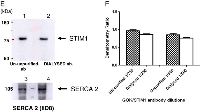
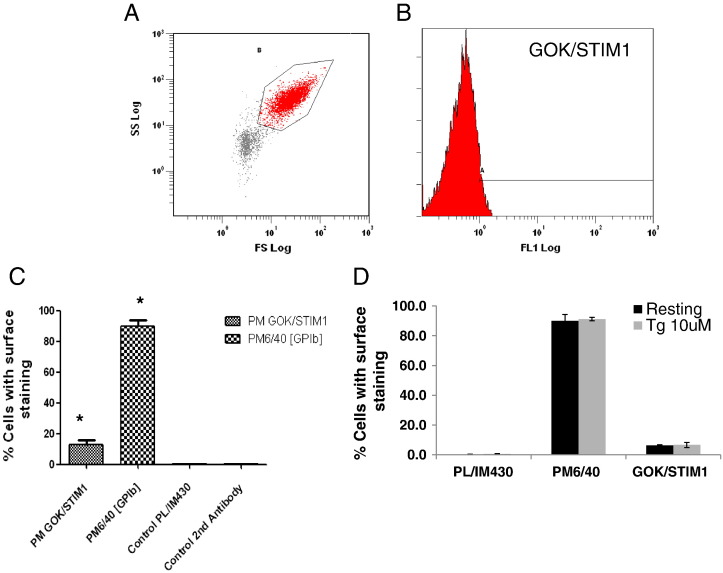
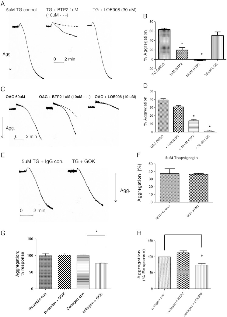
Ca(2+) elevation is essential to platelet activation. STIM1 senses Ca(2+) in the endoplasmic reticulum and activates Orai channels allowing store-operated Ca(2+) entry (SOCE). STIM1 has also been reported to be present in the plasma membrane (PM) with its N-terminal region exposed to the outside medium but its role is not fully understood. We have examined the effects of the antibody GOK/STIM1, which recognises the N-terminal region of STIM1, on SOCE, agonist-stimulated Ca(2+) entry, surface exposure, in vitro thrombus formation and aggregation in human platelets. We also determined novel binding partners of STIM1 using proteomics. The dialysed GOK/STIM1 antibody failed to reduced thapsigargin- and agonist-mediated Ca(2+) entry in Fura2-labelled cells. Using flow cytometry we detect a portion of STIM1 to be surface-exposed. The dialysed GOK/STIM1 antibody reduced thrombus formation by whole blood on collagen-coated capillaries under flow and platelet aggregation induced by collagen. In immunoprecipitation experiments followed by proteomic analysis, STIM1 was found to extract a number of proteins including myosin, DOCK10, thrombospondin-1 and actin. These studies suggest that PM STIM1 may facilitate platelet activation by collagen through novel interactions at the plasma membrane while the essential Ca(2+)-sensing role of STIM1 is served by the protein in the ER.

Keywords: 1,2-diacyl-sn-glycerol; 1-Oleoyl-2-acetyl-sn-glycerol; Aggregation; Ca(2+) entry; Collagen; DAG; OAG; PM; SOCE; STIM1; TG; TRPC; Thrombospondin-1; plasma membrane; store operated Ca(2+) entry; stromal interaction molecule 1; thapsigargin; transient receptor potential canonical.


Figures

Fig. 1
Fig. 1
Purified GOK/STIM1 antibody does not inhibit Ca2 + entry in human platelets. Fura2 labelled platelets were incubated with purified GOK/STIM1 or control PBS at 5 μg/ml for 30 min followed by analysis of Ca2 + elevation using 340/380 nm ratio fluorescence. A. Platelets stimulated with thrombin (at 1.25 U/ml) in the presence of added 100 μM EGTA followed by 1 mM Ca2 + addition to measure Ca2 + entry. Black lines indicate incubation of platelets with control PBS, red lines with dialysed GOK/STIM1 antibody and blue lines in the presence of 0.006% sodium azide. Dotted lines in A and B indicate recordings in the absence of agonist addition but including EGTA and Ca2 +. Typically peak ratio increase in Ca2 + entry for thrombin in the presence of PBS = 0.749 ± 0.004 [n = 3], in the presence of dialysed GOK/STIM1 = 0.714 ± 0.032 [n = 3; P = 0.382], in the presence of sodium azide = 0.461 ± 0.076 [n = 3, P = 0.038; ٭]. B. Responses in platelets stimulated by 1 μg/ml convulxin (CvX). With Ca2 + release differences between the peak height were not significant (P = 0.09). Increase of ratio value for Ca2 + entry in the presence of PBS = 0.57 ± 0.062 [n = 3], in the presence of dialysed GOK/STIM1 = 0.543 ± 0.041 [n = 3, P = 0.06; not significant], in the presence of sodium azide = 0.323 ± 0.031 [n = 3, P = 0.011; ٭٭]. C. Platelets stimulated by 3 μM TG. D. Ca2 + entry expressed as % ratio peak heights after Ca2 + addition for each agonist. Unfilled black bars reflect PBS control, red bars in the presence of GOK/STIM1 antibody and blue bars in the presence of sodium azide. *P = 0.03; **P = 0.01; ***P = 0.001 all compared to controls. Values are from means ± SEM (n = 3). E. Western blotting analysis of STIM1 in platelet lysates using un-purified GOK/STIM1 antibody (lane 1; 1/250 dilution) and dialysed GOK/STIM1 antibody (lane 2; 1/250 dilution). All lanes had 50 μg protein; lanes 3 and 4 were probed with SERCA 2 antibody IID8 (1/2000 dilution). F. Analysis of STIM1 recognition in platelet lysates by 2 dilutions of un-purified and dialysed GOK/STIM1 antibodies by western blotting. Detected bands were scanned by densitometry and expressed as a ratio of the recognition of SERCA 2 by IID8. P values were not significant.
Fig. 1
Fig. 1
Purified GOK/STIM1 antibody does not inhibit Ca2 + entry in human platelets. Fura2 labelled platelets were incubated with purified GOK/STIM1 or control PBS at 5 μg/ml for 30 min followed by analysis of Ca2 + elevation using 340/380 nm ratio fluorescence. A. Platelets stimulated with thrombin (at 1.25 U/ml) in the presence of added 100 μM EGTA followed by 1 mM Ca2 + addition to measure Ca2 + entry. Black lines indicate incubation of platelets with control PBS, red lines with dialysed GOK/STIM1 antibody and blue lines in the presence of 0.006% sodium azide. Dotted lines in A and B indicate recordings in the absence of agonist addition but including EGTA and Ca2 +. Typically peak ratio increase in Ca2 + entry for thrombin in the presence of PBS = 0.749 ± 0.004 [n = 3], in the presence of dialysed GOK/STIM1 = 0.714 ± 0.032 [n = 3; P = 0.382], in the presence of sodium azide = 0.461 ± 0.076 [n = 3, P = 0.038; ٭]. B. Responses in platelets stimulated by 1 μg/ml convulxin (CvX). With Ca2 + release differences between the peak height were not significant (P = 0.09). Increase of ratio value for Ca2 + entry in the presence of PBS = 0.57 ± 0.062 [n = 3], in the presence of dialysed GOK/STIM1 = 0.543 ± 0.041 [n = 3, P = 0.06; not significant], in the presence of sodium azide = 0.323 ± 0.031 [n = 3, P = 0.011; ٭٭]. C. Platelets stimulated by 3 μM TG. D. Ca2 + entry expressed as % ratio peak heights after Ca2 + addition for each agonist. Unfilled black bars reflect PBS control, red bars in the presence of GOK/STIM1 antibody and blue bars in the presence of sodium azide. *P = 0.03; **P = 0.01; ***P = 0.001 all compared to controls. Values are from means ± SEM (n = 3). E. Western blotting analysis of STIM1 in platelet lysates using un-purified GOK/STIM1 antibody (lane 1; 1/250 dilution) and dialysed GOK/STIM1 antibody (lane 2; 1/250 dilution). All lanes had 50 μg protein; lanes 3 and 4 were probed with SERCA 2 antibody IID8 (1/2000 dilution). F. Analysis of STIM1 recognition in platelet lysates by 2 dilutions of un-purified and dialysed GOK/STIM1 antibodies by western blotting. Detected bands were scanned by densitometry and expressed as a ratio of the recognition of SERCA 2 by IID8. P values were not significant.
Fig. 2
Fig. 2
Surface expression of STIM1 using flow cytometry. Platelets were incubated with primary antibodies followed by goat anti-mouse antibody conjugated to Alexa Fluor 488 (2nd antibody). After fixation labelled cells were analysed by flow cytometry counting 10,000 events. A. Scatter plot showing gating of platelets. Particles outside of the gated area were detected in the absence of cells. B. Detection of cells labelled with GOK/STIM1 antibody (% cells stained 12.7% ± 3.2%; P = 0.0026) vs control second antibody (% cells stained 0.4% ± 0.15, P = 0.025) vs PL/IM430 (% cells stained 0.5% ± 0.06). C. Bar chart showing % cells with surface staining by respective antibodies. *P = 0.002 compared to secondary antibody control. D. Bar chart showing effects of TG treatment (10 μM) on surface expression of STIM1 (GOK/STIM1), GP1B (PM6/40) and SERCA 3 (PL/IM430). No significance was noted between TG treatments for each set (n = 3).
Fig. 3
Fig. 3
GOK/STIM1 reduces thrombus formation on collagen coated capillary under flow. Upper panel, representative images of thrombi formed on collagen coated capillaries using blood incubated with control purified antibody (PL/IM430; upper left), dialysed GOK/STIM1 antibody (centre) and dialysed GP1B antibody (PM6/40) (upper right). Bar chart, analysis of mean thrombus volume after 4 min perfusion at a shear rate of 1000 s− 1 (GOK/STIM1 vs control P = 0.048; n = 5; PM6/40 vs control P = 0.028; n = 3).
Fig. 4
Fig. 4
Effects of BTP2, LOE908 and the dialysed GOK/STIM1 antibody on platelet aggregation. For BTP2 and LOE908 studies the inhibitor (or vehicle) was added 5 min prior to addition of agonist (TG, OAG etc.) with 1 mM Ca2 + added 3 min prior to agonist. A + B, BTP2 or LOE908 at indicated doses on TG induced platelet aggregation. C + D, BTP2 or LOE908 on OAG induced platelet aggregation. E + F, Effects of dialysed antibodies at 10 μg/ml for 30 min (GOK/STIM1 [GOK] or IgG control) on TG induced platelet aggregation. G, Effects of GOK/STIM1 on collagen (10 μg/ml) and thrombin (0.5 U/ml) induced platelet aggregation. H, Effects of BTP2 or LOE908 on collagen (15 μg/ml) induced platelet aggregation. Where shown traces are typical of six determinations. Arrow down reflects increased light transmission (aggregation) set with buffer at 100% and platelet suspension at 0%. * reflects P < 0.05.
Fig. 5
Fig. 5
STIM1 does not co-immunoprecipitate TRPC6 or TRPC3 in human platelets. Platelets (1 × 109 cells/ml) were treated with reagents as indicated for 1 min and lysates prepared. Immunoprecipitation was carried out with C terminal polyclonal antibody to STIM1, split into three and subjected to western blotting analysis in parallel. Immunoblots were probed with GOK/STIM1 (1/250 dilution), anti-TRPC6 (SL-TC6; 1/500 dilution) and anti-TRPC3 (A1978; 1/200 dilution) antibodies. QBI + hTRPC3 and QBI − hTRPC6 reflect lysates from hTRPC3 or hTRPC6 over-expressed in QBI-293 cells for positive identification of the respective channels. Blots are typical of three separate determinations.
Fig. 6
Fig. 6
Immunoprecipitation of STIM1 for proteomic analysis and western blots. A. STIM1 was immunoprecipitated using an antibody recognising the C terminus of STIM1 from TG treated platelets (1 min) with the SDS-PAGE gels stained by Colloidal Coomassie stain (TG pla. Lane 3). Lane 1 mm = molecular markers; lane 2 Ab con = antibody reagent control including protein G; lane 3 STIM1 immunoprecipitate. Arrows reflect prominent bands immunoprecipitated by the STIM1 antibody. Bands and clear areas of the gel were used for peptide identification (see Materials and methods). Numbers on the left reflect the size of molecular markers in kDa. B. Co-immunoprecipitation of TSP1 and myosin with STIM1 from platelet lysates. WB-STIM1 probed with GOK/STIM1 (1/250 dilution), TSP-1 (1/200 dilution) and Myosin (1/1000 dilution) antibodies as indicated in Materials and methods. Ab control lane reflects a total reagent control with antibody, protein G etc. but without platelets.

Similar articles

Cited by

References

    1. Authi K.S. Handb. Exp. Pharmacol. 2007;179:425–443. - PubMed
    1. Authi K.S. Blood. 2009;113:1872–1873. - PubMed
    1. Authi K.S., Bokkala S., Patel Y., Kakkar V.V., Munkonge F. Biochem. J. 1993;294(Pt 1):119–126. - PMC - PubMed
    1. Authi K.S., Hughes K., Crawford N. FEBS Lett. 1989;254:52–58. - PubMed
    1. Baba Y., Hayashi K., Fujii Y., Mizushima A., Watarai H., Wakamori M., Numaga T., Mori Y., Iino M., Hikida M., Kurosaki T. Proc. Natl. Acad. Sci. U. S. A. 2006;103:16704–16709. - PMC - PubMed

Publication types

MeSH terms

Substances