Review
. 2014 Mar;26(3):549-55.
doi: 10.1016/j.cellsig.2013.11.028. Epub 2013 Dec 2.

The role of cell signalling in the crosstalk between autophagy and apoptosis

Affiliations
Review

The role of cell signalling in the crosstalk between autophagy and apoptosis

Laurence A Booth et al. Cell Signal. 2014 Mar.

Abstract

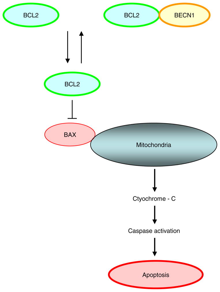
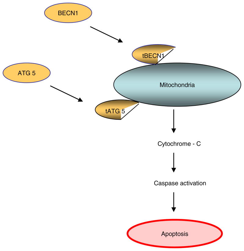
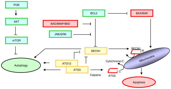
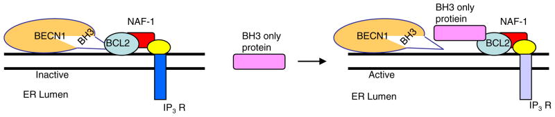
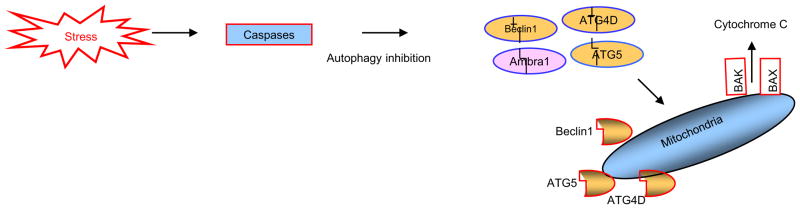
Not surprisingly, the death of a cell is a complex and well controlled process. For several decades, apoptosis, the first genetically programmed death process to be identified has taken centre stage as the principal mechanism of programmed cell death (type I cell death) in mammalian tissues. Apoptosis has been extensively studied and its contribution to the pathogenesis of disease well documented. However, apoptosis does not function alone in determining the fate of a cell. More recently, autophagy, a process in which de novo formed membrane enclosed vesicles engulf and consume cellular components, has been shown to engage in complex interplay with apoptosis. As a result, cell death has been subdivided into the categories apoptosis (Type I), autophagic cell death (Type II), and necrosis (Type III). The boundary between Type I and II cell death is not completely clear and as we will discuss in this review and perhaps a discrete difference does not exist, due to intrinsic factors among different cell types and crosstalk among organelles within each cell type. Apoptosis may begin with autophagy and autophagy can often end with apoptosis, inhibition or a blockade of caspase activity may lead a cell to default into Type II cell death from Type I.

Keywords: Apoptosis; Autophagosome; Autophagy; Caspase; Sorafenib.


Figures

Fig. 1
Fig. 1
BCL2 and BCLXL can inhibit apoptosis by blocking activation of BAX and BAK by preventing subsequent release of cytochrome c and other apoptogenic proteins from the mitochondria.
Fig. 2
Fig. 2
The autophagy regulator ATG5 induces caspase activation following cleavage of Atg 5 by calpain and translocation of the truncated N terminal Atg 5 protein fragment to the outermitichondrial membrane, leading to the induction mitochondrial cytochrome c release and caspase activation. Similarly, translocation of C terminal BECN1 fragment to the outer mitochondrial membrane can induce cytochrome-c release and apoptosis.
Fig. 3
Fig. 3
Phosphorylation of BCL2 at multiple sites by c Jun N terminal protein kinase 1 (JNK1) and extracellular signal related kinase (ERK) have been shown to reduce binding of BCL2 to Beclin 1, leading to the activation of autophagy However, once released from BCL2, as shown in figure 2, translocation of C terminal fragment of BECN1 to the outer mitochondrial membrane can induce cytochrome-c release and apoptosis.
Fig. 4
Fig. 4
Beclin1/BCL2/NAF1 interaction and autophagy regulation at the endo[plasmic reticulum. IP3R—inositol triphosphate receptor.
Fig. 5
Fig. 5
Apoptotic proteins can dismantle the autophagic machinery by degrading Ambra1, Beclin1, ATG4D and ATG5, through the actions of caspases and calpains. Of note is the fact that the proteolytic products of Beclin1 ATG5 and ATG4D may then translocate to the outer mitochondrial membrane and exhibit a pro-apoptotic activity.

Similar articles

Cited by

References

    1. Maiuri MC, Zalckvar E, Kimchi A, Kroemer G. Nat Rev Mol Cell Biol. 2007;8:741–752. - PubMed
    1. Levine B, Yuan J. J Clin Invest. 2005;115:2679–2688. - PMC - PubMed
    1. Lockshin RA, Zahra Z. Int J Biochem Cell Biol. 2004;36:2405–2419. - PubMed
    1. Eisenberg Lerner A, Bialik S, Simon HU, Kimchi A. Cell Death Differ. 2009;16:966–975. - PubMed
    1. White E, DiPaola RS. Clin Cancer Res. 2009;15:5308–5316. - PMC - PubMed

MeSH terms