. 2014 Mar;28(3):1422-34.
doi: 10.1096/fj.13-243840. Epub 2013 Dec 5.

Dopamine D3 receptor inhibits the ubiquitin-specific peptidase 48 to promote NHE3 degradation

Affiliations

Dopamine D3 receptor inhibits the ubiquitin-specific peptidase 48 to promote NHE3 degradation

Ines Armando et al. FASEB J. 2014 Mar.

Abstract

The dopamine D3 receptor (D3R) is crucial in the regulation of blood pressure and sodium balance, in that Drd3 gene ablation in mice results in hypertension and failure to excrete a dietary salt load. The mechanism responsible for the renal sodium retention in these mice is largely unknown. We now offer and describe a novel mechanism by which D3R decreases sodium transport in the long term by inhibiting the deubiquitinylating activity of ubiquitin-specific peptidase 48 (USP48), thereby promoting Na(+)-H(+) exchanger (NHE)-3 degradation. We found that stimulation with the D3R-specific agonist PD128907 (1 μM, 30 min) promoted the interaction and colocalization among D3R, NHE3, and USP48; inhibited USP48 activity (-35±6%, vs. vehicle), resulting in increased ubiquitinylated NHE3 (+140±10%); and decreased NHE3 expression (-50±9%) in human renal proximal tubule cells (hRPTCs). USP48 silencing decreased NHE3's half-life (USP48 siRNA t1/2=6.1 h vs. vehicle t1/2=12.9 h), whereas overexpression of USP48 increased NHE3 half-life (t1/2=21.8 h), indicating that USP48 protects NHE3 from degradation via deubiquitinylation. USP48 accounted for ∼30% of the total deubiquitinylating activity in these cells. Extending our studies in vivo, we found that pharmacologic blockade of D3R via the D3R-specific antagonist GR103691 (1 μg/kg/min, 4 d) in C57Bl/6J mice increased renal NHE3 expression (+310±15%, vs. vehicle), whereas an innovative kidney-restricted Usp48 silencing via siRNA (3 μg/d, 7 d) increased ubiquitinylated NHE3 (+250±30%, vs. controls), decreased total NHE3 (-23±2%), and lowered blood pressure (-24±2 mm Hg), compared with that in control mice that received either the vehicle or nonsilencing siRNA. Our data demonstrate a crucial role for the dynamic interaction between D3R and USP48 in the regulation of NHE3 expression and function.

Keywords: C57Bl/6 mice; blood pressure; kidney; renal proximal tubule cells.


Figures

Figure 1.
Figure 1.
D3R interaction with NHE3. A) Cellular distribution and colocalization of D3R and NHE3 in hRPTCs treated with the D3R agonist PD128907 (1 μM) at the indicated time points. Images were obtained via laser scanning confocal microscope. D3R was pseudocolored green, and NHE3 was pseudocolored red. Colocalization was observed as discrete yellow areas in merged images. WGA tagged with Alexa Fluor 647 was used to target the lectins on the plasma membrane and DAPI to visualize the nucleus. All experiments were performed 3 times (×600; scale bar=10 μm). B) Top: uniform amounts of the lysate and immunoprecipitating antibody were used for the coimmunoprecipitation of D3R and NHE3 in hRPTCs treated with the D3R agonist PD128907 (1 μM) at the indicated time points. NC, negative control (normal mouse IgG); PC, positive control (regular immunoblot of hRPTC lysate). *P < 0.05 vs. basal; 1-way ANOVA and Holm-Sidak post hoc test (dn=3–4). Bottom: coimmunoprecipitation of NHE3 and D3R in hRPTCs treated with PD128907 for 30 min; immunoprecipitation and immunoblot data are in reverse order, relative to the data in the top panel. Normal mouse IgG was used as the negative control. C) Effect of 30-min treatment with a D3R agonist (PD128907, 1 μM), D3R antagonist (GR103691, 1 μM), or both on the coimmunoprecipitation of D3R and NHE3 in hRPTCs. Immunoblots of the D3R and glyceraldehyde 3-phosphate dehydrogenase (GAPDH) from total cell lysates are shown to indicate uniformity of protein loading. *P < 0.05, vs. control (vehicle); 1-way ANOVA and Holm-Sidak post hoc test (n=3). D, E) Abundance of NHE3 in the plasma membrane of hRPTCs treated with the D3R agonist PD128907 (1 μM/30 min), indicating the extent of NHE3 internalization (D) or degradation in the plasma membrane (E), the latter in cells that were incubated in 0.4 M sucrose to inhibit endocytosis. Actin was used for normalization. *P < 0.05 vs. control; Student's t test (n=3). F) Effect of 24-h treatment with quinpirole (Q, a mixed D2R/D3R agonist, 1 μM) in the presence or absence of the specific D3R antagonist GR103691 (GR, 1 μM) or vehicle (V) on the protein abundance of NHE3 in hRPTCs. Molecular sizes: NHE3, 90 kDa; D3R, ∼60 kDa; GAPDH, 35 kDa. *P < 0.05 vs. basal; 1-way ANOVA and Holm-Sidak post hoc test (n=3–4).
Figure 2.
Figure 2.
NHE3 ubiquitinylation and NHE3-Ub distribution. Extent of NHE3 ubiquitinylation (A) and cellular distribution of ubiquitinylated NHE3 (B) in the plasma membrane–enriched fractions and cytoplasm containing intravesicular fractions of hRPTCs in response to the D3R agonist PD128907 (1 μM) at the indicated time points. Abundance of ubiquitinylated NHE3 was determined via coimmunoprecipitation with an anti-Ub antibody as the immunoprecipitant. Immunoblots are shown above the corresponding bar graphs. Molecular sizes: NHE3, 90 kDa; NHE3-Ub, 100 kDa. *P < 0.05 vs. basal; 1-way ANOVA and Holm-Sidak post hoc test (n=3–4).
Figure 3.
Figure 3.
D3R interaction with USP48. A) His pulldown of endogenous USP48 in hRPTCs with heterologously expressed D3R-myc,His. Cells transfected with the empty vector were used as the negative control (n=3/group). Ab, antibody. B) Coimmunoprecipitation of endogenous D3R and USP48 in hRPTCs treated with the D3R agonist PD128907 (1 μM) at the indicated time points. NC, negative control (rabbit mouse IgG), PC, positive control (regular immunoblot of hRPTC lysate). *P < 0.05 vs. basal; 1-way ANOVA and Holm-Sidak post hoc test (n=3). C) Cellular distribution and colocalization of D3R and USP48 in hRPTCs treated with the D3R agonist PD128907 (1 μM) at the indicated time points. Images were obtained via laser scanning confocal microscope. USP48 was pseudocolored red, and D3R was pseudocolored green. Colocalization was observed as discrete yellow areas in merged images. WGA tagged with Alexa Fluor 647 was used to target the lectins on the plasma membrane and DAPI to visualize the nucleus (×600; scale bar=10 μm, n=3). D) Immunoblots of USP48 in hRPTCs depleted of its endogenous USP48 pool and in cells overexpressing USP48. Overexpression was achieved by transfection of pcDNA6.2-V5::hUSP48, whereas depletion of endogenous USP48 was achieved by transfection of USP48-specific siRNA. Actin was used as the housekeeping protein. E, F) USP48 deubiquitinylating activity in hRPTCs in response to stimulation with the D3R agonist PD128907 (1 μM, 30 min; E) or as a function of USP48 expression (overexpression or siRNA-mediated depletion; F). USP48 assays were performed in duplicate. Molecular sizes: USP48, 120 kDa; actin, 40 kDa. *P < 0 .05 vs. basal; 1-way ANOVA and Holm-Sidak post hoc test (n=3). #P < 0 .05 vs. (−) PD128907 treatment; Student's t test (n=3).
Figure 4.
Figure 4.
USP48 interaction with NHE3. A) Distribution of NHE3 and USP48 in lipid raft (fractions 1–6) and non-lipid-raft (fractions 7–12) membrane microdomains of hRPTCs under basal conditions and after treatment with β-MCD (2%), an agent that disrupts lipid rafts by cholesterol depletion. Flotillin-1 and caveolin-1 are lipid raft markers. B) Colocalization of NHE3 and the CTxB, which targets the lipid raft marker ganglioside GM1, in the basal state and after treatment with the D3R agonist PD128907 (1 μM, 15 min). Images were obtained via laser scanning confocal microscope. NHE3 was pseudocolored green, and CTxB was pseudocolored red. Colocalization was observed as discrete yellow areas in merged images (×600, scale bar=10 μm, n=2). C) Cellular distribution and colocalization of NHE3 and USP48 in hRPTCs treated with the D3R agonist PD128907 (1 μM) at the indicated time points. Colocalization was observed as discrete yellow areas in merged images. WGA tagged with Alexa Fluor 647 was used to target the lectins on the plasma membrane and DAPI to visualize the nucleus (×600, scale bar=10 μm, n=3. D) Coimmunoprecipitation of endogenous NHE3 and USP48 in hRPTCs treated with the D3R agonist PD128907 (1 μM) at the indicated time points. An immunoblot is shown above the corresponding bar graph. NC, negative control (rabbit mouse IgG); PC, positive control (regular immunoblot of hRPTC lysate). *P < 0.05 vs. basal; 1-way ANOVA and Holm-Sidak post hoc test (n=3 performed in duplicate). E) Effect of USP48 expression on NHE3 degradation measured using [35S]-Met/Cys labeling. Overexpression was achieved by transfection of pcDNA6.2-V5::hUSP48, whereas depletion of endogenous USP48 was achieved by transfection of USP48-specific siRNA. Radiographs are shown above the line graph. F, G) Total NHE3 and ubiquitinylated NHE3 (NHE3-Ub) in hRPTCs that were either overexpressing (USP48; F) or depleted of USP48 (USP48 siRNA; G). Cells that were transfected with empty vector (EV; F) or nonsilencing siRNA (Mock; G) served as controls. Actin was used for normalization of total NHE3. Molecular sizes: NHE3, 90 kDa; USP48, 120 kDa; Flotillin-1, 50 kDa; Caveolin-1, 25 kDa; Actin, 40 kDa; NHE3-Ub, ∼100 kDa. *P < 0.05 vs. control; Student's t test (n=3).
Figure 5.
Figure 5.
NHE3 localization at the proteasome or lysosome. Cellular distribution and colocalization of NHE3 with the proteasome (A) or lysosome (B) in hRPTCs treated with the D3R agonist PD128907 (1 μM) at the indicated time points. Images were obtained via laser scanning confocal microscope. NHE3 was pseudocolored red; proteasome (via p44S10 marker) or lysosome (via LAMP-1 marker) was pseudocolored red. Colocalization was observed as discrete yellow areas in merged images (×600, scale bar=10 μm, n=3). WGA was used to target the lectins on the plasma membrane and DAPI to visualize the nucleus.
Figure 6.
Figure 6.
Effect of Drd3 deletion on blood pressure and renal NHE3 expression. A) Systolic and diastolic blood pressures of Drd3−/− knockout mice and wild-type littermates on a normal salt diet, measured in animals under pentobarbital anesthesia. B) Renal NHE3 expression in Drd3−/− mice and wild-type littermates on a normal salt diet. *P < 0.05 vs. basal; Student's t test (n=3 performed in duplicate). C) NHE3 immunostaining in renal proximal tubules of Drd3−/− mice and wild-type littermates. Left: ×200. Right: ×1000. PT, proximal tubule; G, glomerulus. D) mRNA profiles of NHE3 in the renal cortices of Drd3−/− mice and wild-type littermates. E) NHE3 and NCC expression profiles in C57Bl/6J mice on a high-salt (1.6% NaCl) diet and treated with the D3R antagonist GR103691 (1 μg/kg/min for 4 d via miniosmotic pump) or vehicle. Molecular sizes: NHE3, 90 kDa; NCC, 115 kDa. *P < 0.05 vs. D3R+/+; 1-way ANOVA and Holm-Sidak post hoc test (n=4/group).
Figure 7.
Figure 7.
Effect of kidney-restricted USP48 depletion on NHE3 and NHE3-Ub abundance and blood pressure in mice. A–C) Expression profiles of endogenous USP48, NHE3, and ubiquitinylated NHE3 (NHE3-Ub) in renal cortices of C57Bl/6J mice that received renal infusion of Usp48-specific siRNA, nonsilencing “mock” siRNA, or vehicle through an osmotic minipump. GAPDH was used for normalization. C) NHE3-Ub was visualized by immunoprecipitating with anti-NHE3 antibody and immunoblotting for Ub. Immunoblots are shown above the corresponding bar graphs. *P < 0.05 vs. vehicle; 1-way ANOVA and Holm-Sidak post hoc test (n=3–4/group). #P < 0.05 vs. mock siRNA transfection or vehicle; Student's t test (n=3–4/group). D, E) Systolic (D) and diastolic (E) blood pressures of uninephrectomized C67Bl/6J mice that had Usp48-specific siRNA, nonsilencing “mock” siRNA, or vehicle infused into the renal subcapsular space through an osmotic pump. The blood pressure was measured before and after the siRNA infusion. Molecular sizes: USP48, 120 kDa; GSPDH, 35 kDa; NHE3, 90 kDa; NHE3-Ub, 100 kDa.*P < 0.05 vs. other groups; 1-way ANOVA and Holm-Sidak post hoc test (n=3–4/group).
Figure 8.
Figure 8.
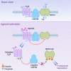
Proposed schema for the dynamic interaction among D3R, USP48, and NHE3 in hRPTCs. D3R, USP48, and NHE3 cofractionate in lipid raft membrane microdomains in the basal state, suggesting the proximity of these key proteins for an effective signal transduction. Ligand occupation of the receptor causes a change in receptor conformation followed by receptor phosphorylation by GRK4 and internalization (31), which effectively attenuates the receptor's responsiveness to subsequent stimulus (receptor desensitization), leading to the internalization of NHE3 and its inactivation by D3R directly, through protein–protein transregulation, or indirectly via PLC and PKC signaling (23) or NHE3 ubiquitinylation (current study). The internalized D3R colocalizes with and inactivates the USP48, thereby preventing the removal of the Ub tags from NHE3-Ub and promoting NHE3-Ub degradation, presumably via the proteasome.

Similar articles

Cited by

References

    1. Felder R. A., Seikaly M. G., Cody P., Eisner G. M., Jose P. A. (1990) Attenuated renal response to dopaminergic drugs in spontaneously hypertensive rats. Hypertension 15, 560–569 - PubMed
    1. Chen C. J., Lokhandwala M. F. (1992) An impairment of renal tubular DA-1 receptor function as the causative factor for diminished natriuresis to volume expansion in spontaneously hypertensive rats. Clin. Exp. Hypertens. A. 14, 615–628 - PubMed
    1. Luippold G., Küster E., Joos T. O., Mühlbauer B. (1998) Dopamine D3 receptor activation modulates renal function in anesthetized rats. Naunyn Schmiedebergs Arch. Pharmacol. 358, 690–693 - PubMed
    1. Ladines C. A., Zeng C., Asico L. D., Sun X., Pocchiari F., Semeraro C., Pisegna J., Wank S., Yamaguchi I., Eisner G. M., Jose P. A. (2001) Impaired renal D1-like and D2-like dopamine receptor interaction in the spontaneously hypertensive rat. Am. J. Physiol. Regul. Integr. Comp. Physiol. 281, R1071–R1078 - PubMed
    1. Sibley D. R. (1999) New insights into dopaminergic receptor function using antisense and genetically altered animals. Annu. Rev. Pharmacol. Toxicol. 39, 313–314 - PubMed

Publication types

LinkOut - more resources