. 2014 Feb;25(2):407-19.
doi: 10.1681/ASN.2013050564. Epub 2013 Dec 5.

The kidney as a reservoir for HIV-1 after renal transplantation

Affiliations

The kidney as a reservoir for HIV-1 after renal transplantation

Guillaume Canaud et al. J Am Soc Nephrol. 2014 Feb.

Abstract

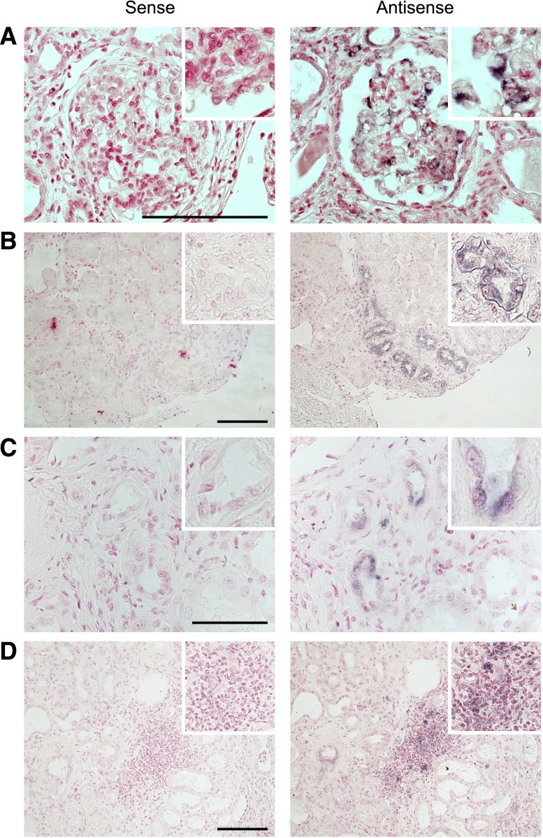
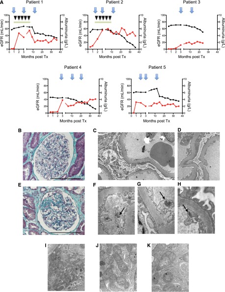
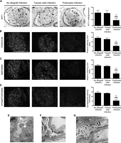
Since the recent publication of data showing favorable outcomes for patients with HIV-1 and ESRD, kidney transplantation has become a therapeutic option in this population. However, reports have documented unexplained reduced allograft survival in these patients. We hypothesized that the unrecognized infection of the transplanted kidney by HIV-1 can compromise long-term allograft function. Using electron microscopy and molecular biology, we examined protocol renal transplant biopsies from 19 recipients with HIV-1 who did not have detectable levels of plasma HIV-1 RNA at transplantation. We found that HIV-1 infected the kidney allograft in 68% of these patients. Notably, HIV-1 infection was detected in either podocytes predominately (38% of recipients) or tubular cells only (62% of recipients). Podocyte infection associated with podocyte apoptosis and loss of differentiation markers as well as a faster decline in allograft function compared with tubular cell infection. In allografts with tubular cell infection, epithelial cells of the proximal convoluted tubules frequently contained abnormal mitochondria, and both patients who developed features of subclinical acute cellular rejection had allografts with tubular cell infection. Finally, we provide a novel noninvasive test for determining HIV-1 infection of the kidney allograft by measuring HIV-1 DNA and RNA levels in patients' urine. In conclusion, HIV-1 can infect kidney allografts after transplantation despite undetectable viremia, and this infection might influence graft outcome.


Figures

Figure 1.
Figure 1.
HIV-1 reinfects the kidney allograft after transplantation. (A) Representative ISH of HIV-1 RNA performed with either sense (negative control) or antisense probes on kidney transplant biopsy performed at the onset of proteinuria. ISH reveals the presence of HIV-1 RNA in podocytes. (B) Representative ISH of HIV-1 RNA performed with either sense (negative control) or antisense probes on kidney transplant biopsy performed at 3 months after transplantation. ISH reveals the presence of HIV-1 RNA in the tubular compartment. (C) Representative ISH of HIV-1 RNA performed with either sense (negative control) or antisense probes on kidney transplant biopsy performed at 3 months after transplantation. ISH reveals the presence of HIV-1 RNA in tubular and vascular sections. (D) Representative ISH of HIV-1 RNA performed with either sense (negative control) or antisense probes on kidney transplant biopsy performed at 3 months after transplantation. ISH reveals the presence of HIV-1 RNA in the inflammatory infiltrates. Infiltrating cells and tubular cells are indistinguishable. Scale bar, 50 μm.
Figure 2.
Figure 2.
Distinct clinical and morphologic features between the two groups of patients. (A) Representative graphics for eGFR (black plot) and albuminuria (red plot) for each patient. The first two patients are treated with plasmapheresis (inverted black triangles) and high-dose cyclosporine (green rectangles). The blue arrows represent transplant biopsies. (B) Representative trichrome staining of glomerular structures at the onset of albuminuria in patients with HIV-1 podocyte infection. The optical microscopy observations are considered normal. (C) Representative electron microscopy image in patients with HIV-1 podocyte infection. Electron microscopy reveals effacement of the foot processes, thickening of the glomerular basement membrane, and vacuolization of the cytoplasm of podocytes. (D) High-power view of the foot process effacement. (E) Representative trichrome staining of the focal and segmental lesions observed in protocol biopsies at 12 months after transplantation in patients with HIV-1 podocyte infection. (F–H) Representative electron microscopy images for patients 1, 2, and 3, respectively. High-power images of a capillary loop showing tubuloreticular inclusions (virus-like particles) within the cytoplasm of endothelial cells (arrows). (I– K) Representative electron microscopy images in patients with HIV-1 tubular cell infection. The epithelial cells of the proximal convoluted tubules frequently contain abnormal mitochondria (i.e., significant variability in size and shape, annulated or distorted cristae, increase in mitochondrial matrix). Scale bar, 100 μm. Original magnification, ×12,000 in C; ×30,000 in D; ×48,000 in F–H; ×15,000 in I; ×53,000 in J; ×70,000 in K. eGFR, estimated GFR; Tx, transplantation.
Figure 3.
Figure 3.
Podocyte infection is associated with podocyte dedifferentiation and depletion at 12 months after transplantation (transplantation). (A) WT1 immunostaining and quantification of WT1-positive glomerular cells in biopsies from HIV-1 transplantation recipients without allograft infection, HIV-1 transplantation recipients with tubular cell infection, and HIV-1 transplantation recipients with podocyte infection at 12 months after transplantation. (B) Podocin immunostaining and quantification of podocin glomerular area in biopsies from HIV-1 transplantation recipients without allograft infection, HIV-1 transplantation recipients with tubular cell infection, and HIV-1 transplantation recipients with podocyte infection at 12 months after transplantation. (C) Nephrin immunostaining and quantification of the nephrin glomerular area in biopsies from HIV-1 transplantation recipients without allograft infection, HIV-1 transplantation recipients with tubular cell infection, and HIV-1 transplantation recipients with podocyte infection at 12 months after transplantation. (D) Synaptopodin immunostaining and quantification of synaptopodin glomerular area in biopsies from HIV-1 transplantation recipients without allograft infection, HIV-1 transplantation recipients with tubular cell infection, and HIV-1 transplantation recipients with podocyte infection at 12 months after transplantation. (E) Representative electron microscopy image in patients with HIV-1 podocyte infection. Low-power view showing slight increase of mesangial matrix, diffuse foot process effacement, and focal podocyte necrosis. (F) High-power field of a necrotic foot process (arrow) between two viable foot processes. (G) Podocytes are necrotic with detachment of foot processes from glomerular basement membrane (arrows). Data are the mean±SD from ANOVA followed by the Tukey-Kramer test. ***P<0.001, HIV-1 transplantation recipients without allograft infection versus HIV-1 transplantation recipients with podocyte infection; ###P<0.001, HIV-1 transplantation recipients with tubular cell infection versus HIV-1 transplantation recipients with podocyte infection. Scale bar, 100 μm. Original magnification, ×2000 in E; ×30,000 in F; ×12,500 in G.

Comment in

References

    1. Adih WK, Selik RM, Hu X: Trends in diseases reported on US death certificates that mentioned HIV infection, 1996-2006. J Int Assoc Physicians AIDS Care (Chic) 10: 5–11, 2011 - PubMed
    1. Wyatt CM, Meliambro K, Klotman PE: Recent progress in HIV-associated nephropathy. Annu Rev Med 63: 147–159, 2012 - PubMed
    1. Cohen MS, Shaw GM, McMichael AJ, Haynes BF: Acute HIV-1 Infection. N Engl J Med 364: 1943–1954, 2011 - PMC - PubMed
    1. Marras D, Bruggeman LA, Gao F, Tanji N, Mansukhani MM, Cara A, Ross MD, Gusella GL, Benson G, D’Agati VD, Hahn BH, Klotman ME, Klotman PE: Replication and compartmentalization of HIV-1 in kidney epithelium of patients with HIV-associated nephropathy. Nat Med 8: 522–526, 2002 - PubMed
    1. Stock PG, Barin B, Murphy B, Hanto D, Diego JM, Light J, Davis C, Blumberg E, Simon D, Subramanian A, Millis JM, Lyon GM, Brayman K, Slakey D, Shapiro R, Melancon J, Jacobson JM, Stosor V, Olson JL, Stablein DM, Roland ME: Outcomes of kidney transplantation in HIV-infected recipients. N Engl J Med 363: 2004–2014, 2010 - PMC - PubMed

MeSH terms