. 2014 Feb;137(Pt 2):420-32.
doi: 10.1093/brain/awt327. Epub 2013 Dec 5.

Neuronal merlin influences ERBB2 receptor expression on Schwann cells through neuregulin 1 type III signalling

Affiliations

Neuronal merlin influences ERBB2 receptor expression on Schwann cells through neuregulin 1 type III signalling

Alexander Schulz et al. Brain. 2014 Feb.

Abstract

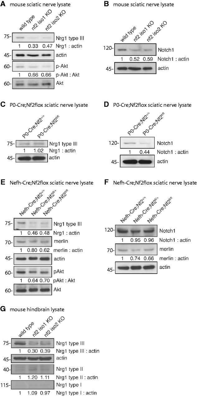
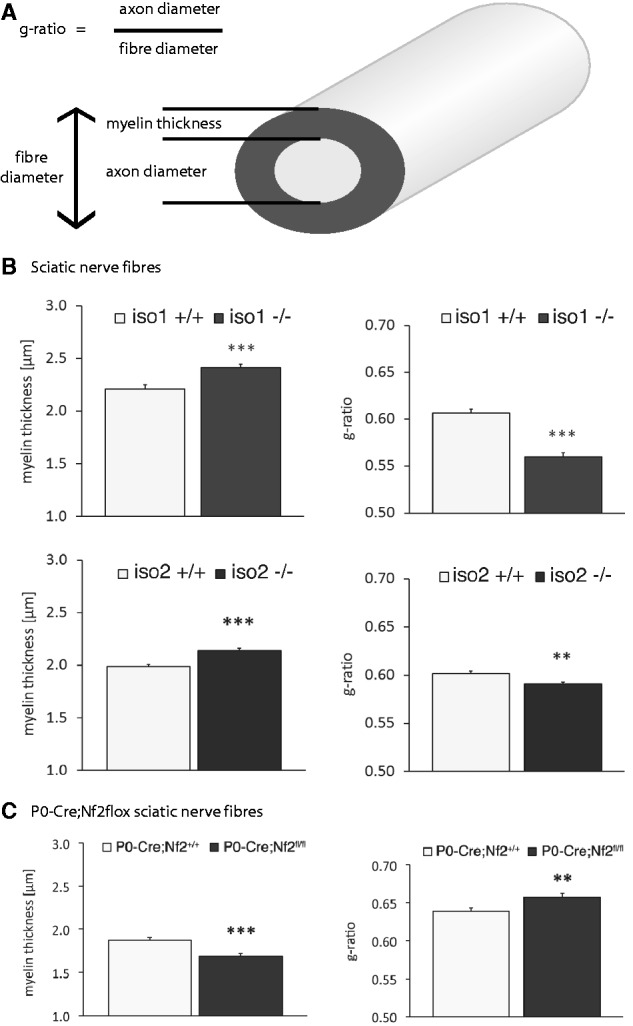
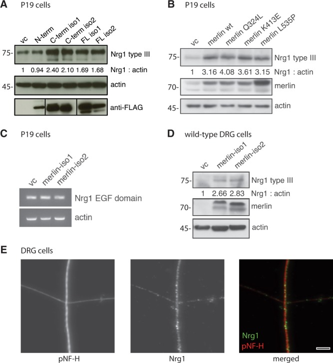
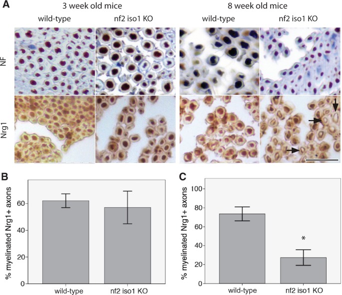
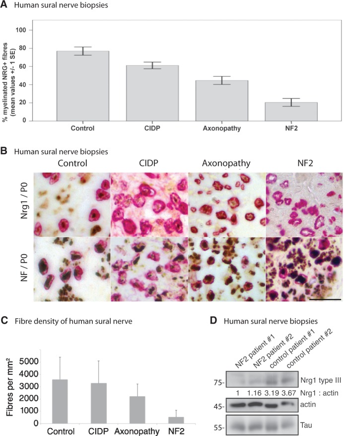
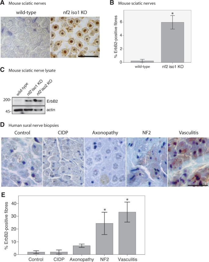
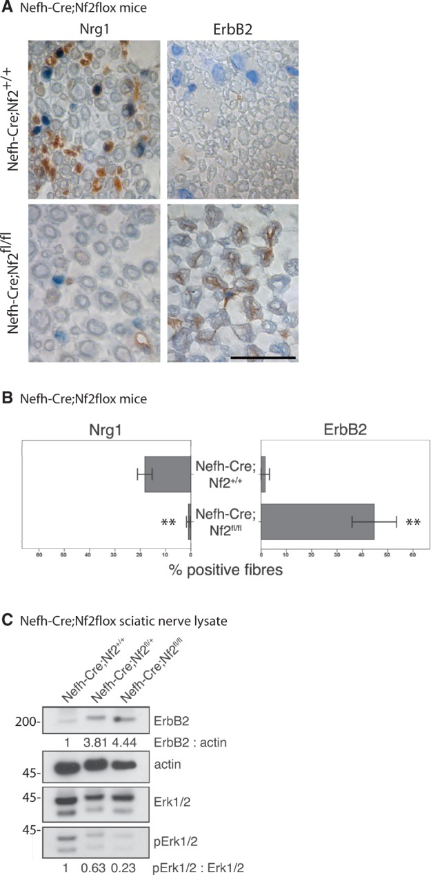
Axonal surface proteins encompass a group of heterogeneous molecules, which exert a variety of different functions in the highly interdependent relationship between axons and Schwann cells. We recently revealed that the tumour suppressor protein merlin, mutated in the hereditary tumour syndrome neurofibromatosis type 2, impacts significantly on axon structure maintenance in the peripheral nervous system. We now report on a role of neuronal merlin in the regulation of the axonal surface protein neuregulin 1 important for modulating Schwann cell differentiation and myelination. Specifically, neuregulin 1 type III expression is reduced in sciatic nerve tissue of neuron-specific knockout animals as well as in biopsies from seven patients with neurofibromatosis type 2. In vitro experiments performed on both the P19 neuronal cell line and primary dorsal root ganglion cells demonstrate the influence of merlin on neuregulin 1 type III expression. Moreover, expression of ERBB2, a Schwann cell receptor for neuregulin 1 ligands is increased in nerve tissue of both neuron-specific merlin knockout animals and patients with neurofibromatosis type 2, demonstrating for the first time that axonal merlin indirectly regulates Schwann cell behaviour. Collectively, we have identified that neuronally expressed merlin can influence Schwann cell activity in a cell-extrinsic manner.

Keywords: Schwann cell; axon; merlin; myelination; neuregulin 1; neurofibromatosis type 2; polyneuropathy.


Figures

Figure 1
Figure 1
Loss of merlin in sciatic nerve lysates is accompanied by decreasing NRG1 type III levels. (A–F) Immunoblots of sciatic nerve lysates taken from adult mice. (A) Loss of each major merlin isoform in transgenic animals (nf2 iso1 KO; nf2 iso2 KO) results in decreased expression of NRG1 type III and reduced Akt phosphorylation (p-Akt). Actin was used as loading control (n = 3). (B) Loss of merlin-iso1 or merlin-iso2 in vivo leads to reduced expression of NOTCH1 (n = 2). (C and D) Schwann cell-specific loss of merlin (P0-Cre;Nf2fl/fl) has no effect on NRG1 type III level but lowers NOTCH1 expression compared with wild-type littermates (P0-Cre;Nf2+/+; n = 3). (E and F) Neuron-specific merlin knockout (Nefh-Cre;Nf2floxfl/fl) reduces levels of NRG1 type III and p-Akt. NOTCH1 levels are unchanged in sciatic nerves after loss of merlin in the neuronal compartment (n = 2). (G) Isoform-specific loss of merlin reduces NRG1 type III amounts in hind-brain lysates of adult mice (n = 3). Blot quantifications (density values) are depicted below respective lanes and are normalized to actin and wild-type controls. KO = knockout.
Figure 2
Figure 2
Merlin knockout animals show altered myelination. (A) Schematic diagram indicating important nerve fibre parameters. (B and C) Myelination thickness and g-ratio quantifications of sciatic nerves taken from (B) merlin isoform-specific knockout animals (iso1 -/-; iso2 -/-) and wild-type littermates (iso1 +/+; iso2 +/+) as well as from (C) P0-Cre;Nf2flox mice bearing a Schwann cell-specific merlin loss (data represent mean ± SEM; **P < 0.01; ***P < 0.001; n > 300).
Figure 3
Figure 3
Merlin overexpression in vitro induces NRG1 type III expression. (A) Immunoblot of P19 cell lysates after transfection of different FLAG-tagged merlin fragments. Transfection of empty vector was used as control (vc); actin indicates equal loading. Anti-FLAG staining shows transfection rate of each construct (n = 3). (B) Wild-type merlin isoform 1 (merlin wt) and merlin constructs bearing indicated C-terminal point mutations were transfected into P19 cells. Immunoblot shows NRG1 type III levels as well as merlin and actin as transfection and loading control, respectively (n = 2). Blot quantifications (density values) are depicted below respective lanes and are normalized to actin and transfection controls (vc). (C) Reverse-transcription PCR of P19 cell lysates following overexpression of merlin-iso1 or merlin-iso2 constructs. EGF domain-specific primers were used to detect Nrg1 transcripts. Actin was used as loading control (n = 3). (D) Immunoblot of lysates derived from primary dorsal root ganglion (DRG) cells after merlin overexpression in vitro (n = 3). Blot quantifications (density values) are depicted below respective lanes and are normalized to actin and transfection controls (vc). (E) Immunostaining of primary dorsal root ganglion cells shows localization of NRG1 (green) in axons of cultured dorsal root ganglions (4 days in vitro). Axons were stained for phosphorylated neurofilaments as an axonal marker (pNF-H, red). Scale bar = 5 µm. N-term = N-terminal fragment; C-Term = C-terminal fragment; FL = full length.
Figure 4
Figure 4
Sciatic nerve axons of merlin knockout mice show reduced axonal NRG1 expression. (A) Representative pictures of immunohistochemical stainings for neurofilaments (NF) and NRG1 on sciatic nerves of merlin isoform1-deficient mice (nf2 iso1 KO) and wild-type controls. Visualization of bound antibodies with diaminobenzidine (brown), counterstained with Mayer’s haemalum. Scale bar = 20 µm. Arrows indicate examples of unstained axons. (B and C) Percentage of NRG1-positive myelinated fibres in sciatic nerve samples of 3-week-old (B) and 8-week-old (C) nf2 iso1 knockout (KO) mice and corresponding wild-type littermates (data represent mean ± SEM; *P < 0.05).
Figure 5
Figure 5
Sural nerve biopsies of NF2 patients show reduced NRG1 type III levels. (A) Percentage of NRG1-positive myelinated fibres in nerve samples of healthy control individuals versus chronic inflammatory demyelinating polyneuropathy (CIDP), axonopathy and patients with NF2 (data represent mean ± SEM; for significance levels see ‘Results’ section). (B) Expression of NRG1/myelin protein zero (P0) versus neurofilament (NF)/myelin protein zero in sural nerve biopsies of healthy control individuals as well as chronic inflammatory demyelinating polyneuropathy, axonopathy and patients with NF2. Typical examples of immunohistochemical double-labelling. Visualization of bound neurofilament (NF) and NRG1 antibodies with diaminobenzidine (brown) and of myelin protein zero antibodies with fast red (red), counterstained with Mayer’s haemalum. Scale bar = 20 µm. (C) Sural nerve fibre densities per mm2 of indicated disease conditions (data represent mean ± SEM). (D) Sural nerve biopsies of patients with NF2 and healthy control subjects were lysed and immunoblotted for NRG1 type III. Actin and the axonal marker tau served as loading control (n = 3). Blot quantifications (density values) are depicted below respective lanes and are normalized to actin and healthy control samples.
Figure 6
Figure 6
ERBB2 expression is upregulated in sural nerve biopsies of patients with NF2. (A) Representative examples of immunohistochemical stainings for ERBB2 on mouse sciatic nerves deficient of merlin isoform 1 (nf2 iso1 KO) and wild-type controls. Visualization of bound antibodies with diaminobenzidine (brown), counterstained with Mayer’s haemalum. Scale bar = 20 µm. (B) Related quantifications to (A) of ERBB2-positive fibres in wild-type mice and merlin isoform1 knockout animals (data represent mean ± SEM; *P < 0.05; n = 3). (C) Immunoblot of sciatic nerve lysates showing that the loss of both major merlin isoforms in vivo is accompanied by increased ERBB2 levels (n = 2). Blot quantifications (density values) are depicted below respective lanes and are normalized to actin and wild-type samples. (D) Representative examples of immunohistochemical labelling of human sural nerve biopsies for ERBB2. Visualization of bound antibodies with diaminobenzidine (brown), counterstained with Mayer’s haemalum. Scale bar = 20 µm. (E) Quantifications of ERBB2-positive fibres in biopsies of controls as well as patients suffering from chronic inflammatory demyelinating polyneuropathy (CIDP), axonopathy, NF2 and vasculitis (data represent mean ± SEM; *P < 0.05; n = 3).
Figure 7
Figure 7
Neuron-specific loss of merlin is sufficient to increase ErbB2 levels on Schwann cells. (A) Representative pictures of immunohistochemical staining of mouse sciatic nerves taken from neuron-specific merlin knockout animals (Nefh-Cre;Nf2fl/fl) and wild-type littermates (Nefh-Cre;Nf2+/+). Visualization of antibodies raised against NRG1 and ERBB2 with diaminobenzidine (brown), counterstained with Mayer's haemalum. Scale bar = 20 µm. (B) Quantifications of NRG1 and ERBB2-positively labelled fibres in neuron-specific merlin knockout mice (Nefh-Cre;Nf2fl/fl) and controls (data represent mean ± SEM, **P < 0.01; n = 3). (C) Immunoblot of sciatic nerve lysates derived from homozygous (Nefh-Cre;Nf2fl/fl) and heterozygous neuron-specific merlin knockout mice (Nefh-Cre;Nf2fl/+) indicates elevated ERBB2 protein levels and decreased phosphorylation of Erk (pErk) compared with wild-type littermates (Nefh-Cre;Nf2+/+; n = 3).

Similar articles

Cited by

References

    1. Baser MER, Evans DG, Gutmann DH. Neurofibromatosis 2. Curr Opin Neurol. 2003;16:27–33. - PubMed
    1. Clark JJ, Provenzano M, Diggelmann HR, Xu N, Hansen SS, Hansen MR. The ErbB inhibitors trastuzumab and erlotinib inhibit growth of vestibular schwannoma xenografts in nude mice: a preliminary study. Otol Neurotol. 2008;29:846–53. - PMC - PubMed
    1. Corfas G, Velardez MO, Ko CP, Ratner N, Peles E. Mechanisms and roles of axon-Schwann cell interactions. J Neurosci. 2004;24:9250–60. - PMC - PubMed
    1. El Bejjani R, Hammarlund M. Notch signaling inhibits axon regeneration. Neuron. 2012;73:268–78. - PMC - PubMed
    1. Fricker FR, Bennett DL. The role of neuregulin-1 in the response to nerve injury. Future Neurol. 2011;6:809–22. - PMC - PubMed

Publication types

MeSH terms