

How vascular endothelial growth factor-A (VEGF) regulates differentiation of mesenchymal stem cells
- PMID: 24309509
- PMCID: PMC3902099
- DOI: 10.1369/0022155413516347
How vascular endothelial growth factor-A (VEGF) regulates differentiation of mesenchymal stem cells
Abstract
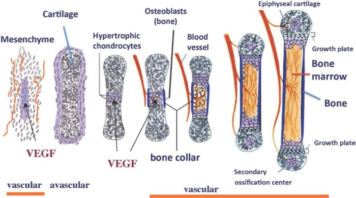
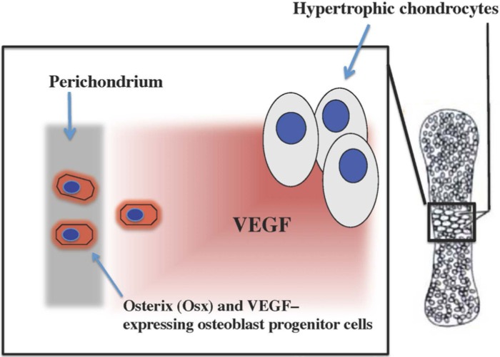
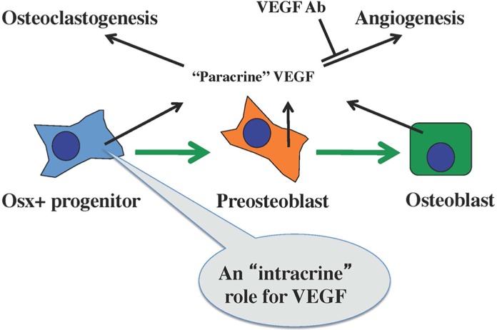
Vascular endothelial growth factor A (VEGF), a key factor in angiogenesis, plays an essential role in skeletal development and postnatal homeostasis. VEGF serves as a survival factor for chondrocytes and couples the resorption of cartilage with bone formation during endochondral ossification. Recently, it has also been found to regulate the balance between osteoblast and adipocyte differentiation in bone marrow mesenchymal stem cells. Surprisingly, this regulatory function of VEGF is not based on paracrine signaling involving cell surface receptor activation. Instead, the mechanism appears to utilize intracellular VEGF, which is functionally linked to the nuclear envelope protein lamin A. Lamin A and VEGF control osteoblast and adipocyte differentiation by regulating the levels of the osteoblast and adipocyte transcription factors Runx2 and PPARγ, respectively. These data raise the intriguing possibility that loss of bone mass during aging may be manipulated by controlling the levels and activity of intracellular VEGF in bone marrow mesenchymal stem cells.
Keywords: VEGF; bone remodeling; osteoblast; skeletal development; transcription.
Conflict of interest statement
Figures
References
-
- Barleon B, Sozzani S, Zhou D, Weich HA, Mantovani A, Marme D. (1996). Migration of human monocytes in response to vascular endothelial growth factor (VEGF) is mediated via the VEGF receptor flt-1. Blood 87:3336-3343 - PubMed
-
- Carmeliet P, Collen D. (1999). Role of vascular endothelial growth factor and vascular endothelial growth factor receptors in vascular development. Curr Top Microbiol Immunol 237:133-158 - PubMed
-
- Carmeliet P, Ferreira V, Breier G, Pollefeyt S, Kieckens L, Gertsenstein M, Fahrig M, Vandenhoeck A, Harpal K, Eberhardt C, Declercq C, Pawling J, Moons L, Collen D, Risau W, Nagy A. (1996). Abnormal blood vessel development and lethality in embryos lacking a single VEGF allele. Nature 380:435-439 - PubMed
-
- de Vries C, Escobedo JA, Ueno H, Houck K, Ferrara N, Williams LT. (1992). The fms-like tyrosine kinase, a receptor for vascular endothelial growth factor. Science 255:989-991 - PubMed
-
- Ferrara N, Carver-Moore K, Chen H, Dowd M, Lu L, O’Shea KS, Powell-Braxton L, Hillan KJ, Moore MW. (1996). Heterozygous embryonic lethality induced by targeted inactivation of the VEGF gene. Nature 380:439-442 - PubMed
Publication types
MeSH terms
Substances
Grants and funding
LinkOut - more resources
Full Text Sources
Other Literature Sources
