. 2013 Dec 5;4(12):e948.
doi: 10.1038/cddis.2013.467.

Low-dose triptolide in combination with idarubicin induces apoptosis in AML leukemic stem-like KG1a cell line by modulation of the intrinsic and extrinsic factors

Affiliations

Low-dose triptolide in combination with idarubicin induces apoptosis in AML leukemic stem-like KG1a cell line by modulation of the intrinsic and extrinsic factors

Y Liu et al. Cell Death Dis. .

Abstract

Leukemia stem cells (LSCs) are considered to be the main reason for relapse and are also regarded as a major hurdle for the success of acute myeloid leukemia chemotherapy. Thus, new drugs targeting LSCs are urgently needed. Triptolide (TPL) is cytotoxic to LSCs. Low dose of TPL enhances the cytotoxicity of idarubicin (IDA) in LSCs. In this study, the ability of TPL to induce apoptosis in leukemic stem cell (LSC)-like cells derived from acute myeloid leukemia cell line KG1a was investigated. LSC-like cells sorted from KG1a were subjected to cell cycle analysis and different treatments, and then followed by in vitro methyl thiazole tetrazolium bromide cytotoxicity assay. The effects of different drug combinations on cell viability, intracellular reactive-oxygen species (ROS) activity, colony-forming ability and apoptotic status were also examined. Combination index-isobologram analysis indicates a synergistic effect between TPL and IDA, which inhibits the colony-forming ability of LSC-like cells and induces their apoptosis. We further investigated the expression of Nrf2, HIF-1α and their downstream target genes. LSC-like cells treated with both TPL and IDA have increased levels of ROS, decreased expression of Nrf2 and HIF-1α pathways. Our findings indicate that the synergistic cytotoxicity of TPL and IDA in LSCs-like cells may attribute to both induction of ROS and inhibition of the Nrf2 and HIF-1α pathways.


Figures

Figure 1
Figure 1
Structure of triptolide
Figure 2
Figure 2
Leukemia stem-like cells were sorted from KG1a cell line. (a) Phenotypes of sorted cells were analyzed using flow cytometry. Before sorting, the CD34+CD38− KG1a cells were 53.24±2.11%. After sorting, the percentage of CD34+CD38− cells is 98.15±1.64%. (b) Cell cycle parameters in the sorted cells were analyzed with G0/G1 cells being 82.4±3.82%. Figures represent three independent experiments
Figure 3
Figure 3
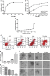
TPL enhances cytotoxicity of IDA to leukemia stem-like cells. (a) Leukemia stem-like cells were exposed to indicated concentrations of TPL for 72 h and then the viability was determined by MTT assay. (b) Cytotoxicity of IDA was determined after exposure to indicated concentrations of IDA with or without TPL (IC20, 5 nM). (c) Synergy was determined by combination index (CI) analysis and expressed as CI versus fraction affected. (d) Leukemia stem-like cells were exposed to different treatments (IDA: 27 nM, TPL: 5 nM) with apoptotic ratio detected using flow cytometry. (e) Histogram of apoptotic ratio in leukemia stem-like cells. (f) Colonies were formed from leukemia stem-like cells being exposed to IDA (27 nM) with or without TPL (5 nM) for 24 h. (g) The colony-forming units grown by leukemia stem-like cells were counted (40 or more cells per unit) per well. All treated groups have decreased colony units with the largest scale of decrease occurring in combined group. TPL, triptolide; IDA, idarubicin; values are expressed as mean±S.D. of three independent experiments
Figure 4
Figure 4
TPL in combination with IDA generates ROS in leukemia stem-like cells. Leukemia stem-like cells were incubated with indicated concentrations of IDA with or without TPL (5 nM) for 24 h. The DCF fluorescence was detected as described in Methods. TPL intensively synergized IDA in inducing ROS activity in leukemia stem-like cells in a concentration-dependent manner. TPL, triptolide; IDA, idarubicin; values are expressed as mean±S.D. of three independent experiments.
Figure 5
Figure 5
TPL in combination of IDA decreases the expression of Nrf2, HIF-1α and its downstream genes. (a) Nrf2 and its downstream genes were assessed using RT-PCR after leukemia stem-like cells being exposed to indicated drugs (IDA, 27 nM plus TPL, 5 nM) for 24 h. RT-PCR result showed that the expression of Nrf2 and NQO1, GSR and HO-1 genes decreased in single-drug-treated groups while the largest scale of decrease occurred in leukemia stem-like cells exposed to IDA plus TPL. (b) HIF-1α and its downstream genes were assessed using RT-PCR after leukemia stem-like cells being exposed to indicated drugs (IDA, 27 nM plus TPL, 5 nM) for 24 h. RT-PCR result shows that the expression of HIF-1α and BNIP3, VEGF and CAIX genes decreased in single-drug-treated groups while the largest scale of decrease occurred in leukemia stem-like cells exposed to IDA plus TPL. (c) Western blotting analysis of the expression of Nrf2, HIF-1α, CXCR4 and VLA-4 in the leukemia stem-like cells treated as indicated. Nrf2, HIF-1α, CXCR4 and VLA-4 were assessed using western blotting analysis after leukemia stem-like cells being exposed to indicated drugs (IDA, 27 nM plus TPL, 5 nM) for 24 h. The expression of Nrf2, HIF-1α, CXCR4 and VLA-4 decreased in treated groups especially in cells treated by IDA+TPL group. GAPDH was used as a loading control. This figure represents three independent experiments. TPL: triptolide; IDA: idarubicin; values are expressed as mean±S.D. of three independent experiments
Figure 6
Figure 6
Mechanism of TPL-induced apoptosis in leukemia stem-like cells. A relatively low concentration of TPL in combination with IDA could induce apoptosis of leukemia stem-like cells in vitro through influence of extrinsic components, for example, downregulation of HIF-1α, CXCR4 and VLA-4 as well as intrinsic components, for example, increase of ROS level and decrease of Nrf2

References

    1. Damiani D, Tiribelli M, Raspadori D, Michelutti A, Gozzetti A, Calistri E, et al. The role of MDR-related proteins in the prognosis of adult acute myeloid leukaemia (AML) with normal karyotype. Hematol Oncol. 2007;25:38–43. - PubMed
    1. Lapidot T, Grunberger T, Vormoor J, Estrov Z, Kollet O, Bunin N, et al. Identification of human juvenile chronic myelogenous leukemia stem cells capable of initiating the disease in primary and secondary SCID mice. Blood. 1996;88:2655–2664. - PubMed
    1. de Figueiredo-Pontes LL, Pintao MC, Oliveira LC, Dalmazzo LF, Jacomo RH, Garcia AB, et al. Determination of P-glycoprotein, MDR-related protein 1, breast cancer resistance protein, and lung-resistance protein expression in leukemic stem cells of acute myeloid leukemia. Cytometry B Clin Cytom. 2008;74:163–168. - PubMed
    1. Fardel O, Payen L, Courtois A, Drenou B, Fauchet R, Rault B. Differential expression and activity of P-glycoprotein and multidrug resistance-associated protein in CD34-positive KG1a leukemic cells. Int J Oncol. 1998;12:315–319. - PubMed
    1. She M, Niu X, Chen X, Li J, Zhou M, He Y, et al. Resistance of leukemic stem-like cells in AML cell line KG1a to natural killer cell-mediated cytotoxicity. Cancer Letters. 2012;318:173–179. - PubMed

Publication types

MeSH terms