

Long-term cultured mesenchymal stem cells frequently develop genomic mutations but do not undergo malignant transformation
- PMID: 24309937
- PMCID: PMC3877551
- DOI: 10.1038/cddis.2013.480
Long-term cultured mesenchymal stem cells frequently develop genomic mutations but do not undergo malignant transformation
Abstract
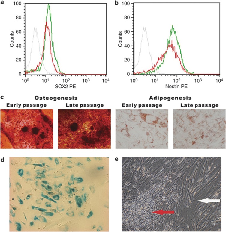
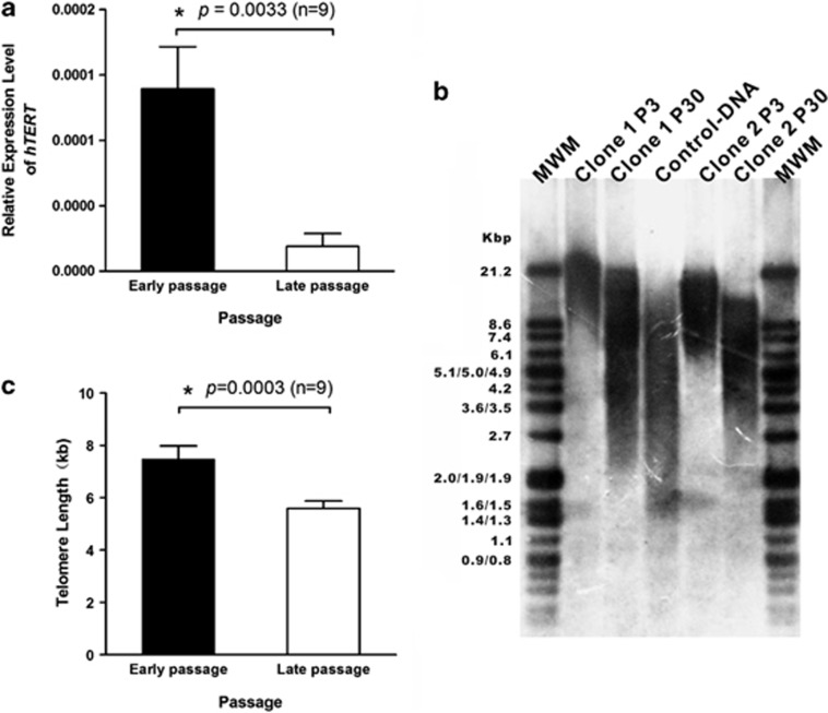
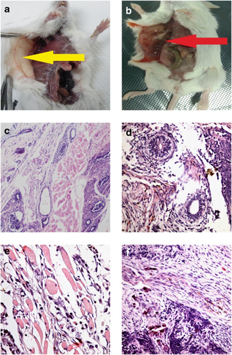
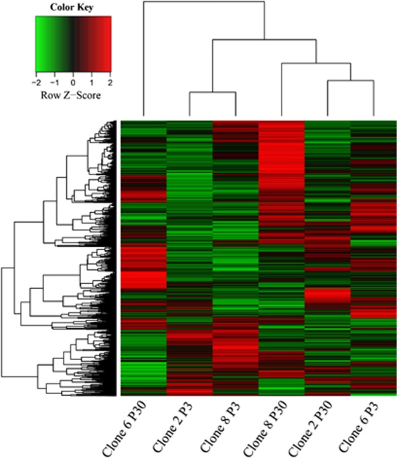
Cultured human umbilical cord mesenchymal stem cells (hUC-MSCs) are being tested in several clinical trials and encouraging outcomes have been observed. To determine whether in vitro expansion influences the genomic stability of hUC-MSCs, we maintained nine hUC-MSC clones in long-term culture and comparatively analyzed them at early and late passages. All of the clones senesced in culture, exhibiting decreased telomerase activity and shortened telomeres. Two clones showed no DNA copy number variations (CNVs) at passage 30 (P30). Seven clones had ≥1 CNVs at P30 compared with P3, and one of these clones appeared trisomic chromosome 10 at the late passage. No tumor developed in immunodeficient mice injected with hUC-MSCs, regardless of whether the cells had CNVs at the late passage. mRNA-Seq analysis indicated that pathways of cell cycle control and DNA damage response were downregulated during in vitro culture in hUC-MSC clones that showed genomic instability, but the same pathways were upregulated in the clones with good genomic stability. These results demonstrated that hUC-MSCs can be cultured for many passages and attain a large number of cells, but most of the cultured hUC-MSCs develop genomic alterations. Although hUC-MSCs with genomic alterations do not undergo malignant transformation, periodic genomic monitoring and donor management focusing on genomic stability are recommended before these cells are used for clinical applications.
Figures
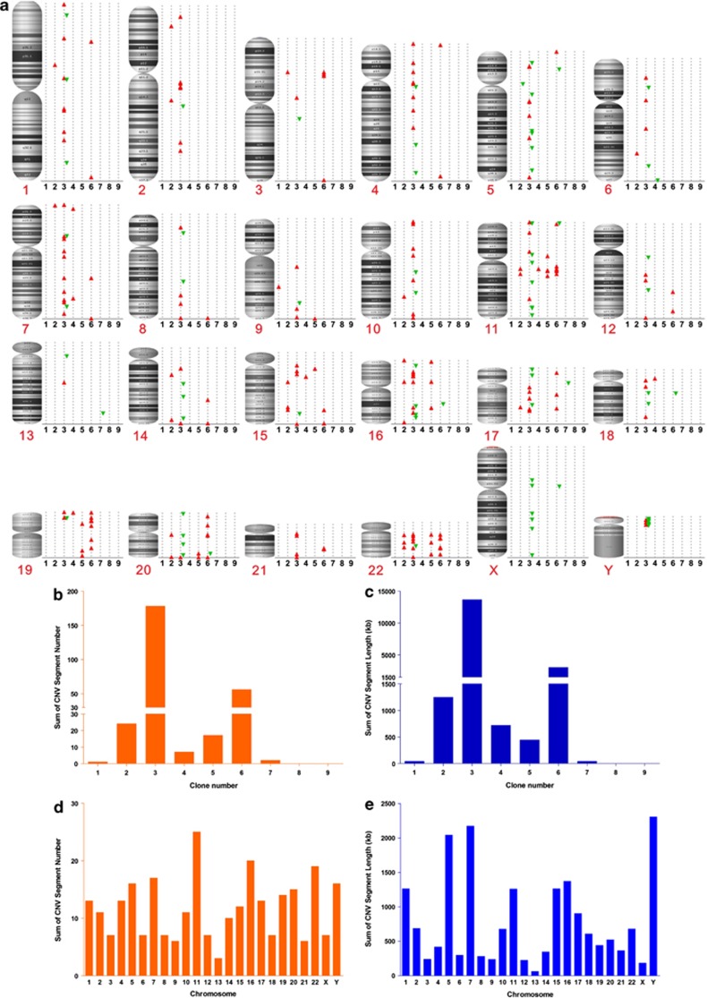
 , amplification;
  , deletion. CNV segment number and length for each hUC-MSC clone are presented in panels b and c, respectively. CNV segment number and length for each chromosome are shown in panels d and e, respectively
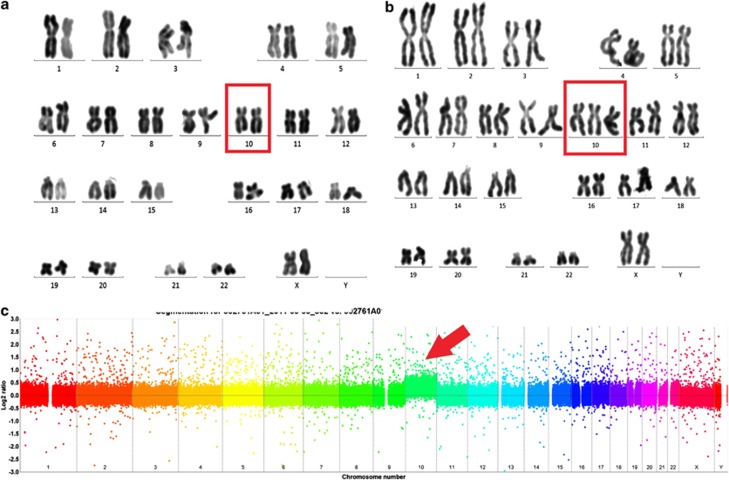
References
-
- Maitra A, Arking DE, Shivapurkar N, Ikeda M, Stastny V, Kassauei K, et al. Genomic alterations in cultured human embryonic stem cells. Nat Genet. 2005;37:1099–1103. - PubMed
-
- Narva E, Autio R, Rahkonen N, Kong L, Harrison N, Kitsberg D, et al. High-resolution DNA analysis of human embryonic stem cell lines reveals culture-induced copy number changes and loss of heterozygosity. Nat Biotechnol. 2010;28:371–377. - PubMed
-
- Lu LL, Liu YJ, Yang SG, Zhao QJ, Wang X, Gong W, et al. Isolation and characterization of human umbilical cord mesenchymal stem cells with hematopoiesis-supportive function and other potentials. Haematologica. 2006;91:1017–1026. - PubMed
Publication types
MeSH terms
LinkOut - more resources
Full Text Sources
Other Literature Sources
Medical
