Review
. 2013 Dec 6;45(12):e66.
doi: 10.1038/emm.2013.97.

Recognition of lipopolysaccharide pattern by TLR4 complexes

Affiliations
Review

Recognition of lipopolysaccharide pattern by TLR4 complexes

Beom Seok Park et al. Exp Mol Med. .

Abstract

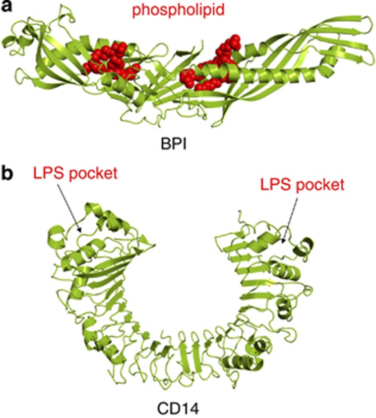
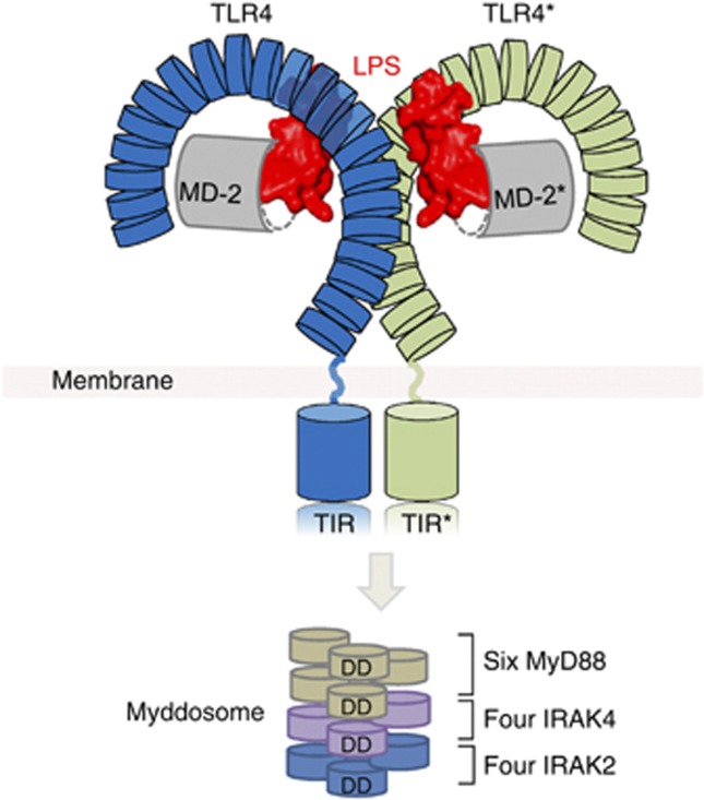
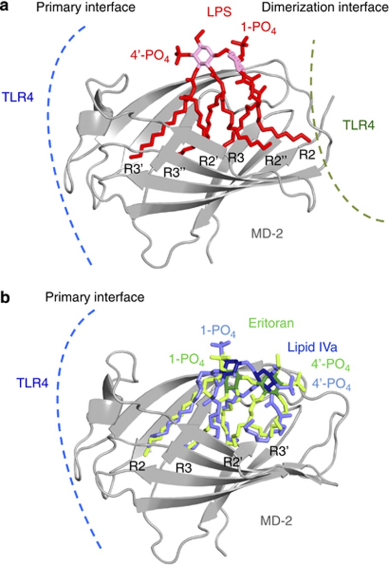
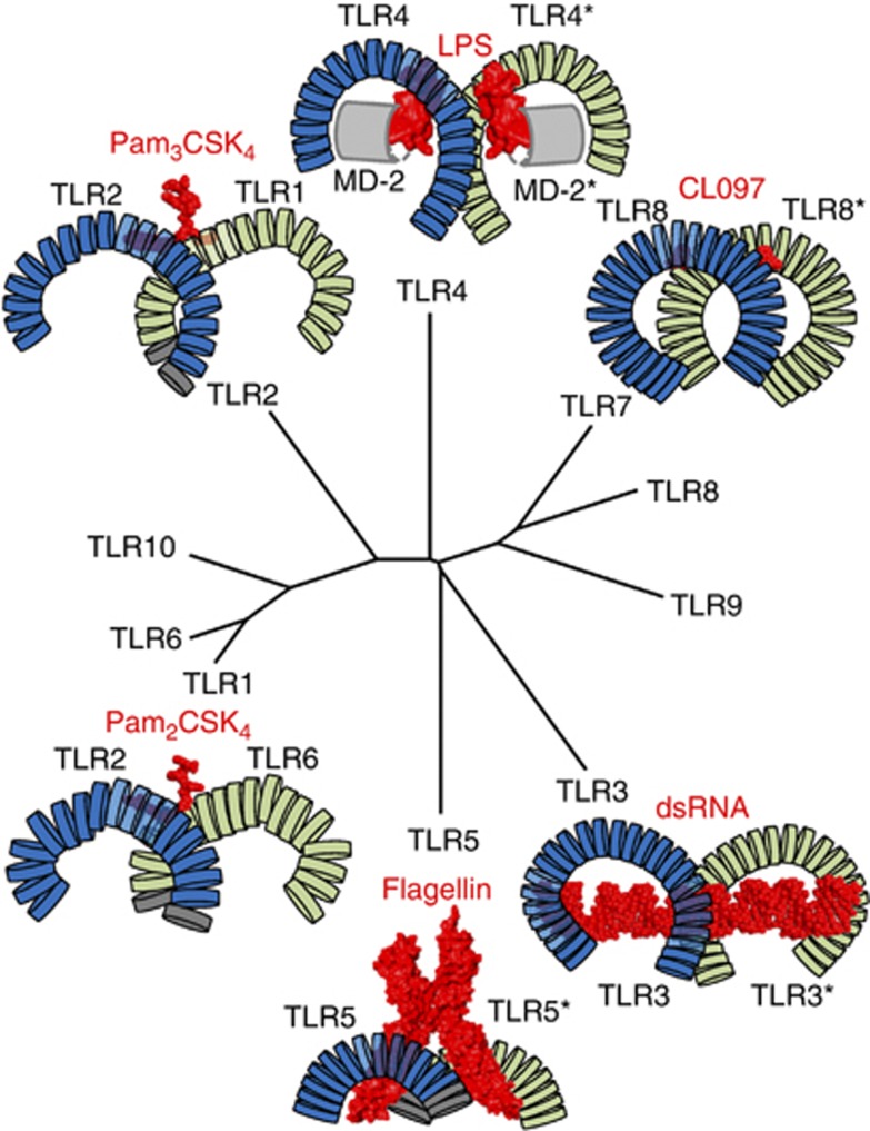
Lipopolysaccharide (LPS) is a major component of the outer membrane of Gram-negative bacteria. Minute amounts of LPS released from infecting pathogens can initiate potent innate immune responses that prime the immune system against further infection. However, when the LPS response is not properly controlled it can lead to fatal septic shock syndrome. The common structural pattern of LPS in diverse bacterial species is recognized by a cascade of LPS receptors and accessory proteins, LPS binding protein (LBP), CD14 and the Toll-like receptor4 (TLR4)-MD-2 complex. The structures of these proteins account for how our immune system differentiates LPS molecules from structurally similar host molecules. They also provide insights useful for discovery of anti-sepsis drugs. In this review, we summarize these structures and describe the structural basis of LPS recognition by LPS receptors and accessory proteins.


Figures

Figure 1
Figure 1
The structures of accessory proteins involved in LPS recognition. (a) The crystal structure of BPI, with two phospholipid binding sites. LBP is expected to have a similar structure. (b) CD14 forms homodimers. The monomeric subunit of CD14 contains 11 LRR modules and a single LRRNT module.
Figure 2
Figure 2
Overview of LPS recognition by TLR4–MD-2. LPS binding induces dimerization of the TLR4–MD-2 complex, which is proposed to enable dimerization of the intracellular TIR domains and recruitment of adaptor molecules such as MyD88. Aggregation of the death domains (DD) of MyD88 brings four IRAK4 and four IRAK2 molecules together forming a large tower-like structure called the ‘Myddosome'.
Figure 3
Figure 3
Binding of LPS and antagonistic ligands to the TLR4–MD-2 complex. (a) Structure of the primary and dimerization interfaces of the TLR4–MD-2–LPS complex. The lipid chains of LPS are labeled. MD-2 is colored grey. The lipid chains and phosphate groups of LPS are shown in red. The glucosamine backbone is pink. (b) Structures of Eritoran and lipid IVa bound to MD-2.
Figure 4
Figure 4
TIR domain structures. (a) The figure was drawn using coordinate files with PDB codes as follows: 1FYV (TLR1), 1FYW (TLR2), 2J67 (TLR10), 2Z5V (MyD88) and 2Y92 (Mal). The backbones of the TIR domains are yellow; positive charges are blue and negative charges red. (b) Sequence alignment of TIR domains. The BB and DD loops important in TLR signaling are boxed in red.
Figure 5
Figure 5
Structures and a phylogenetic tree of TLRs. Crystal structures of TLR4–MD-2–LPS, TLR2–TLR1–Pam3CSK4, TLR2–TLR6–Pam2CSK4, TLR5-flagellin, TLR3-dsRNA, TLR8-CL097 are shown. The ligands are colored red, and TLRs are blue and green.
Figure 6
Figure 6
The hybrid LRR technique. Two LRR family proteins are fused together while maintaining the sequence conservation pattern of the LRR modules. (a) The functional domain of TLR4 fused with a partner LRR protein at its C-terminus. (b) Alternatively, the partner protein can be fused to the N-terminal region of TLR4.

References

    1. Janeway CA, Jr, Medzhitov R. Innate immune recognition. Annu Rev Immunol. 2002;20:197–216. - PubMed
    1. Medzhitov R, Janeway C., Jr Innate immune recognition: mechanisms and pathways. Immunol Rev. 2000;173:89–97. - PubMed
    1. Raetz CR, Whitfield C. Lipopolysaccharide endotoxins. Annu Rev Biochem. 2002;71:635–700. - PMC - PubMed
    1. Pugin J, Schurer-Maly CC, Leturcq D, Moriarty A, Ulevitch RJ, Tobias PS. Lipopolysaccharide activation of human endothelial and epithelial cells is mediated by lipopolysaccharide-binding protein and soluble CD14. Proc Natl Acad Sci USA. 1993;90:2744–2748. - PMC - PubMed
    1. Chow JC, Young DW, Golenbock DT, Christ WJ, Gusovsky F. Toll-like receptor-4 mediates lipopolysaccharide-induced signal transduction. J Biol Chem. 1999;274:10689–10692. - PubMed

Publication types