

Cell lineage tracing reveals a biliary origin of intrahepatic cholangiocarcinoma
- PMID: 24310400
- PMCID: PMC3929349
- DOI: 10.1158/0008-5472.CAN-13-1911
Cell lineage tracing reveals a biliary origin of intrahepatic cholangiocarcinoma
Abstract
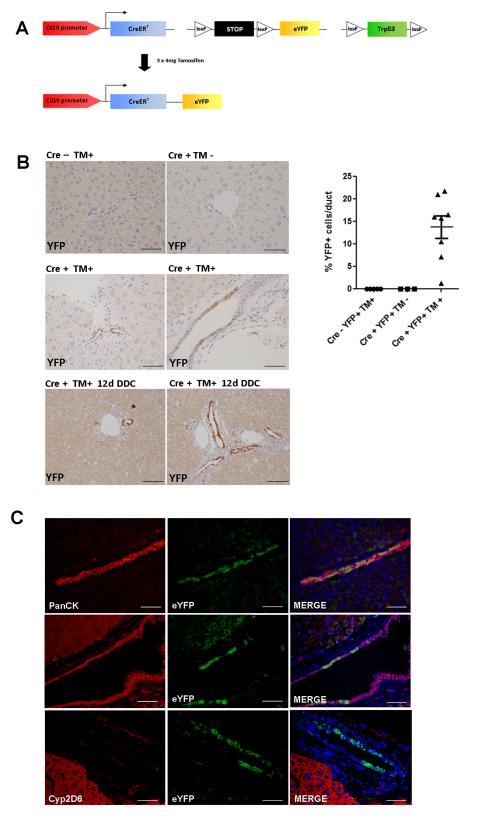
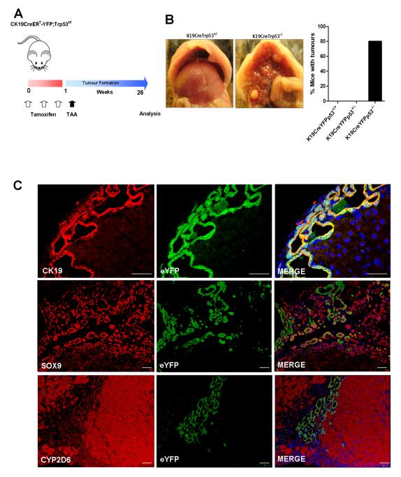
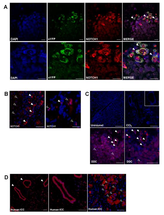
Intrahepatic cholangiocarcinoma is a treatment refractory malignancy with a high mortality and an increasing incidence worldwide. Recent studies have observed that activation of Notch and AKT signaling within mature hepatocytes is able to induce the formation of tumors displaying biliary lineage markers, thereby raising the suggestion that it is hepatocytes, rather than cholangiocytes or hepatic progenitor cells that represent the cell of origin of this tumor. Here, we use a cholangiocyte-lineage tracing system to target p53 loss to biliary epithelia and observe the appearance of labeled biliary lineage tumors in response to chronic injury. Consequent to this, upregulation of native functional Notch signaling is observed to occur spontaneously within cholangiocytes and hepatocytes in this model as well as in human intrahepatic cholangiocarcinoma. These data prove that in the context of chronic inflammation and p53 loss, frequent occurrences in human disease, biliary epithelia are a target of transformation and an origin of intrahepatic cholangiocarcinoma.
©2013 AACR.
Figures
Similar articles
-
Intrahepatic cholangiocarcinoma can arise from Notch-mediated conversion of hepatocytes.J Clin Invest. 2012 Nov;122(11):3914-8. doi: 10.1172/JCI63065. J Clin Invest. 2012. PMID: 23023701 Free PMC article.
-
Kras and Tp53 Mutations Cause Cholangiocyte- and Hepatocyte-Derived Cholangiocarcinoma.Cancer Res. 2018 Aug 15;78(16):4445-4451. doi: 10.1158/0008-5472.CAN-17-1123. Epub 2018 Jun 5. Cancer Res. 2018. PMID: 29871934 Free PMC article.
-
Kupffer cells induce Notch-mediated hepatocyte conversion in a common mouse model of intrahepatic cholangiocarcinoma.Sci Rep. 2016 Oct 4;6:34691. doi: 10.1038/srep34691. Sci Rep. 2016. PMID: 27698452 Free PMC article.
-
Liver stem cells and their implication in hepatocellular and cholangiocarcinoma.Oncogene. 2006 Jun 26;25(27):3818-22. doi: 10.1038/sj.onc.1209558. Oncogene. 2006. PMID: 16799623 Review.
-
YAP1 activation and Hippo pathway signaling in the pathogenesis and treatment of intrahepatic cholangiocarcinoma.Adv Cancer Res. 2022;156:283-317. doi: 10.1016/bs.acr.2022.02.003. Epub 2022 Mar 9. Adv Cancer Res. 2022. PMID: 35961703 Free PMC article. Review.
Cited by
-
Resolving tumor evolution: a phylogenetic approach.J Natl Cancer Cent. 2024 Mar 21;4(2):97-106. doi: 10.1016/j.jncc.2024.03.001. eCollection 2024 Jun. J Natl Cancer Cent. 2024. PMID: 39282584 Free PMC article. Review.
-
Kupffer Cell-Derived Tnf Triggers Cholangiocellular Tumorigenesis through JNK due to Chronic Mitochondrial Dysfunction and ROS.Cancer Cell. 2017 Jun 12;31(6):771-789.e6. doi: 10.1016/j.ccell.2017.05.006. Cancer Cell. 2017. PMID: 28609656 Free PMC article.
-
A novel mouse model of intrahepatic cholangiocarcinoma induced by liver-specific Kras activation and Pten deletion.Sci Rep. 2016 Apr 1;6:23899. doi: 10.1038/srep23899. Sci Rep. 2016. PMID: 27032374 Free PMC article.
-
The potential role of liver stem cells in initiation of primary liver cancer.Hepatol Int. 2016 Nov;10(6):893-901. doi: 10.1007/s12072-016-9730-9. Epub 2016 May 2. Hepatol Int. 2016. PMID: 27139191 Review.
-
Interplay of Cardiometabolic Syndrome and Biliary Tract Cancer: A Comprehensive Analysis with Gender-Specific Insights.Cancers (Basel). 2024 Oct 9;16(19):3432. doi: 10.3390/cancers16193432. Cancers (Basel). 2024. PMID: 39410050 Free PMC article. Review.
References
-
- Ishak KGAP, Sobin LH. Histological typing of tumours of the liver. WHO International Classification of Tumour. Springer Verlag; Berlin: 1994.
-
- Bergquist AEA, Olsson R, Kornfeldt D, Loof L, Danielsson A, Hultcrantz R, Lindgren S, Prytz H, Sandberg-Gertzen H, Almer S, Granath F, Broome U. Hepatic and extrahepatic malignancies in primary sclerosing cholangitis. Journal of Hepatology. 2002;36:321–7. - PubMed
Publication types
MeSH terms
Substances
Grants and funding
LinkOut - more resources
Full Text Sources
Other Literature Sources
Medical
Molecular Biology Databases
Research Materials
Miscellaneous
