. 2013 Dec;69(Pt 12):2555-62.
doi: 10.1107/S0907444913027042. Epub 2013 Nov 19.

Structure of human apurinic/apyrimidinic endonuclease 1 with the essential Mg2+ cofactor

Affiliations

Structure of human apurinic/apyrimidinic endonuclease 1 with the essential Mg2+ cofactor

Brittney A Manvilla et al. Acta Crystallogr D Biol Crystallogr. 2013 Dec.

Abstract

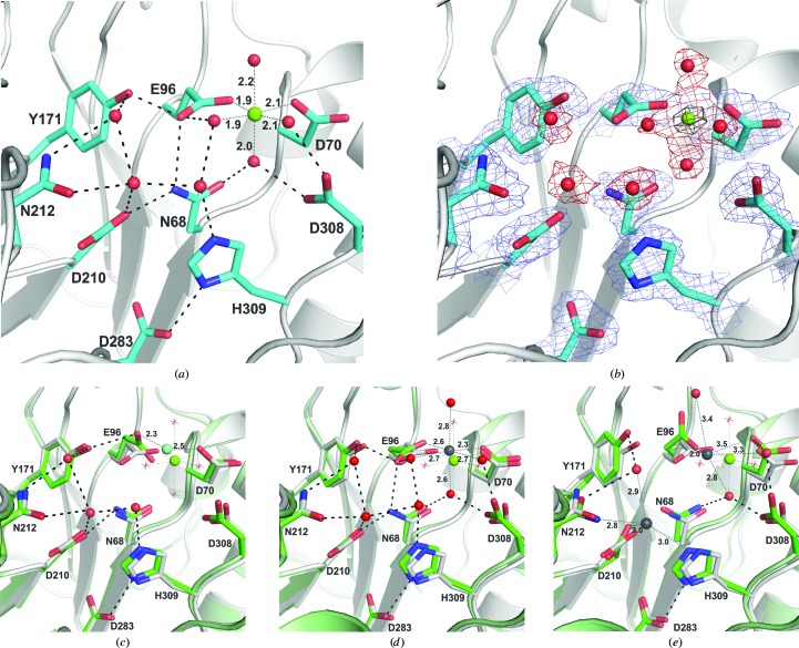
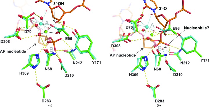
Apurinic/apyrimidinic endonuclease 1 (APE1) mediates the repair of abasic sites and other DNA lesions and is essential for base-excision repair and strand-break repair pathways. APE1 hydrolyzes the phosphodiester bond at abasic sites, producing 5'-deoxyribose phosphate and the 3'-OH primer needed for repair synthesis. It also has additional repair activities, including the removal of 3'-blocking groups. APE1 is a powerful enzyme that absolutely requires Mg2+, but the stoichiometry and catalytic function of the divalent cation remain unresolved for APE1 and for other enzymes in the DNase I superfamily. Previously reported structures of DNA-free APE1 contained either Sm3+ or Pb2+ in the active site. However, these are poor surrogates for Mg2+ because Sm3+ is not a cofactor and Pb2+ inhibits APE1, and their coordination geometry is expected to differ from that of Mg2+. A crystal structure of human APE1 was solved at 1.92 Å resolution with a single Mg2+ ion in the active site. The structure reveals ideal octahedral coordination of Mg2+ via two carboxylate groups and four water molecules. One residue that coordinates Mg2+ directly and two that bind inner-sphere water molecules are strictly conserved in the DNase I superfamily. This structure, together with a recent structure of the enzyme-product complex, inform on the stoichiometry and the role of Mg2+ in APE1-catalyzed reactions.

Keywords: apurinic/apyrimidinic DNA; base-excision repair; nucleases; phosphoryl transfer.


Figures

Figure 1
Figure 1
New crystal structure of human APE1 with the native Mg2+ cofactor and previous structures with surrogate metals. (a) Close-up view of the active site of the new structure, showing Mg2+ and several ordered water molecules (molecule A of the three molecules in the asymmetric unit for PDB entry 4lnd). Also shown are the important catalytic residues; all but Asp70 are strictly conserved in the DNase I superfamily (Tyr171 is replaced by His in some members). The octahedral coordination of Mg2+ is indicated by dotted lines with distances provided (also given in Table 3 ▶). Hydrogen bonds are indicated by dashed lines. (b) The same view of the active site showing a 2F oF c OMIT map contoured at 1.5σ for protein and waters and an F oF c OMIT map contoured at 8.0σ for the Mg2+ ion (black mesh). Note that one or more of the three non-Mg2+-coordinating water molecules are not observed in the other two protein molecules (B and C) in the asymmetric unit. (c) Previously reported structure of Sm3+-bound APE1 (green; PDB entry 1bix; Gorman et al., 1997 ▶) aligned with the new Mg2+-bound structure (white). The coordination of the Sm3+ ion (cyan) is shown (dotted lines) with distances. The coordination of Mg2+ (green) in the new structure is also indicated (without distances). Water molecules shown as red spheres and hydrogen bonds (dashed lines) are for the Sm3+-bound structure. The water molecules that coordinate Mg2+ are shown as red stars. (d) The previously reported structure of APE1 with one Pb2+ ion (green; PDB entry 1hd7; Beernink et al., 2001 ▶) aligned with the new Mg2+-bound structure (white). The coordination of the Pb2+ ion (gray) is shown (dotted lines) with distances and the coordination of Mg2+ (green) is also indicated. Water molecules (red spheres) and hydrogen bonds (dashed lines) are for the Pb2+-bound structure (waters that coordinate Mg2+ are shown as red stars). (e) The previously reported structure of APE1 with two Pb2+ ions (green; PDB entry 1e9n; Beernink et al., 2001 ▶) aligned with the new Mg2+-bound structure (white). The coordination of the Pb2+ ions (gray) is shown (dotted lines) with distances and the coordination of Mg2+ (green) is similarly indicated. Water molecules (red spheres) and hydrogen bonds (dashed lines) are for the Pb2+-bound structure (waters that coordinate Mg2+ are shown as red stars).
Figure 2
Figure 2
Alignment of the new Mg2+-bound APE1 structure with DNA-bound structures. (a) Recently determined structure of the APE1 enzyme–product (EP) complex (green; PDB entry 4iem; Tsutakawa et al., 2013 ▶) aligned with the structure of Mg2+-bound APE1 (cyan) reported here. DNA from the EP complex (orange) contains a 3′-OH and a 5′-deoxyribose phosphate (dRP). The Mg2+ ion from the EP complex is colored green and its coordination is indicated by black dotted lines. The Mg2+ ion from the new DNA-free structure is colored cyan and its coordination is indicated by cyan dotted lines, with coordinating water molecules shown as red stars. Hydrogen-bond interactions (yellow dashes) are shown for the EP complex only. The approximate locations of the A and B sites are noted (gray symbols). (b) Structure of the APE1 enzyme–substrate (ES) complex (green; PDB entry 1dew; Mol et al., 2000 ▶) aligned with the new structure of Mg2+-bound APE1 (cyan). The DNA in the ES complex (orange) contains an intact abasic site and Mg2+ was omitted from the ES complex to halt P—O bond cleavage. The green sphere indicates the position of the Mg2+ ion in the EP complex (also aligned with DNA-free APE1). Mg2+ in the DNA-free structure is shown in cyan; its coordination is indicated by cyan dotted lines, with coordinating waters shown as red stars. Hydrogen-bond interactions (yellow dashes) are shown for the ES complex. The potential nucleophilic water from our DNA-free structure is shown as a red star, with cyan dashes indicating hydrogen bonds to Asp210 and Asn212. This water molecule was also observed in previous structures of APE1 with Sm3+ or a single Pb2+ ion (Fig. 1 ▶). The approximate locations of the A and B sites are noted (gray symbols).

Similar articles

Cited by

References

    1. Abbotts, R. & Madhusudan, S. (2010). Cancer Treat. Rev. 36, 425–435. - PubMed
    1. Adhikari, S., Choudhury, S., Mitra, P. S., Dubash, J. J., Sajankila, S. P. & Roy, R. (2008). Anticancer Agents Med. Chem. 8, 351–357. - PubMed
    1. Barzilay, G., Mol, C. D., Robson, C. N., Walker, L. J., Cunningham, R. P., Tainer, J. A. & Hickson, I. D. (1995). Nature Struct. Biol. 2, 561–568. - PubMed
    1. Beernink, P. T., Segelke, B. W., Hadi, M. Z., Erzberger, J. P., Wilson, D. M. III & Rupp, B. (2001). J. Mol. Biol. 307, 1023–1034. - PubMed
    1. Bhakat, K. K., Mantha, A. K. & Mitra, S. (2009). Antioxid. Redox Signal. 11, 621–638. - PMC - PubMed

Publication types

Associated data