Review
. 2014 Apr;35(2):195-233.
doi: 10.1210/er.2013-1053. Epub 2013 Dec 5.

Clinical and molecular genetics of the phosphodiesterases (PDEs)

Affiliations
Review

Clinical and molecular genetics of the phosphodiesterases (PDEs)

Monalisa F Azevedo et al. Endocr Rev. 2014 Apr.

Abstract

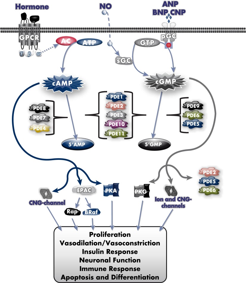
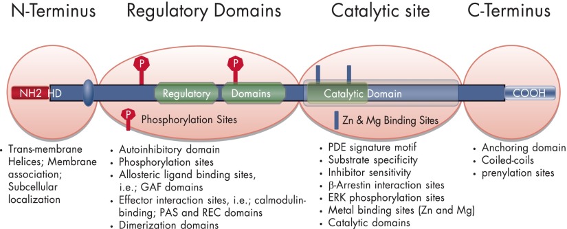
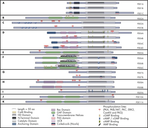
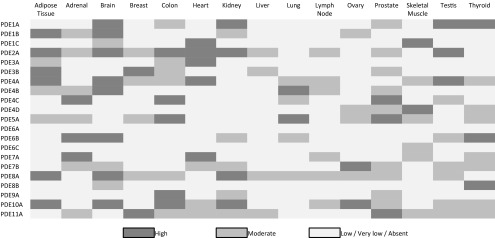
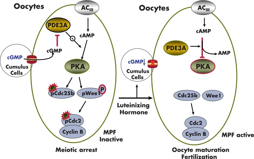
Cyclic nucleotide phosphodiesterases (PDEs) are enzymes that have the unique function of terminating cyclic nucleotide signaling by catalyzing the hydrolysis of cAMP and GMP. They are critical regulators of the intracellular concentrations of cAMP and cGMP as well as of their signaling pathways and downstream biological effects. PDEs have been exploited pharmacologically for more than half a century, and some of the most successful drugs worldwide today affect PDE function. Recently, mutations in PDE genes have been identified as causative of certain human genetic diseases; even more recently, functional variants of PDE genes have been suggested to play a potential role in predisposition to tumors and/or cancer, especially in cAMP-sensitive tissues. Mouse models have been developed that point to wide developmental effects of PDEs from heart function to reproduction, to tumors, and beyond. This review brings together knowledge from a variety of disciplines (biochemistry and pharmacology, oncology, endocrinology, and reproductive sciences) with emphasis on recent research on PDEs, how PDEs affect cAMP and cGMP signaling in health and disease, and what pharmacological exploitations of PDEs may be useful in modulating cyclic nucleotide signaling in a way that prevents or treats certain human diseases.


Figures

Figure 1.
Figure 1.
Cyclic nucleotide signaling and regulation. AC, adenylyl cyclase; ANP, atrial natriuretic peptide; BNP, B-type natriuretic peptide; B-Raf, B-Raf protein kinase; CNG-channel, cyclic nucleotide-gated channel; CNP, C-type natriuretic peptide; pGC, particulate guanylyl cyclase; PKG, cGMP-dependent protein kinase; Rap, Ras-related protein; sAC soluble AC; sGC, soluble guanylyl cyclase.
Figure 2.
Figure 2.
General structure of PDE enzyme molecules. HD, hydrophobic domains.
Figure 3.
Figure 3.
Schematic representation of the structure of each of the 11 human PDE families. HD, hydrophobic domains.
Figure 4.
Figure 4.
PDE tissue expression. Each PDE is considered individually based on its maximum and minimum expression (478–483). Reference: Microarray combined data acquired from the ArrayExpress database (www.ebi.ac.uk/arrayexpress) under the accession numbers E-GEOD-30352, E-AFMX-5, A-AFFY-44, and E-MTAB-513.
Figure 5.
Figure 5.
AKAPs: scaffolds for PDE-containing signalosomes.
Figure 6.
Figure 6.
Myocardial AKAPs. The subcellular localization of different AKAPs is shown, together with their effector proteins. Cav1.2 is regulated by cAMP confined to specific signaling domains. β1- and β2-ARs are associated with Cav1.2 and AKAPs (AKAP79 and AKAP15/18α), which regulate cAMP production and channel activity. AKAP79/150 has a CaN binding domain and is one of the endogenous inhibitors of CaN. Yotiao and KCNQ1 regulate potassium ion current. PDE3A/AKAP18δ/PLB/SERCA2 regulate Ca2+ reuptake. PI3K p110γ/PDE3B complex regulates cAMP and myocardial function. Troponin T, synemin, and myospryn are sarcomeric AKAPs. Synemin is localized at the Z-disk and binds to actinin and desmin to act as a mechanical linker. D-AKAP1 and -2 are localized at the mitochondrial membrane. Expression of AKAP-LBC is critical in the hypertrophic response. mAKAP is specifically localized at, and anchors PKA to, the SR and nuclear membranes. mAKAP interacts with RYR2 and PDE4. AKAP95 is localized at the nuclear membrane, exhibits a cell cycle-dependent interaction with PKA, and coordinates a scaffold of hormonally responsive transcription complexes. A complex of MTG-PKA and PDE7A is shown at Golgi. CaV1.2, LTCCs; KCNQ, IKS potassium channel subunit; SKIP, sphingosine kinase-interacting protein; MTG, myeloid translocation gene; CaN, calcineurin.
Figure 7.
Figure 7.
PDE3A regulates SERCA2-mediated Ca2+ uptake into the SR. A, The diagram illustrates some of the physiological interactions and interplay between two intracellular second messengers, cAMP and calcium, in regulation of the excitation contractile cycle in cardiac myocytes (blue for cAMP; green for calcium) (58, 59). PDE3A regulates SERCA2-mediated Ca2+ uptake into the SR by modulating cAMP/PKA-induced phosphorylation of PLB (pPLB) (395). PLB, an endogenous muscle-specific SERCA2 inhibitor, interacts with SERCA2 and PDE3A and determines the rate of calcium reuptake into the SR, after its release from myofilaments at the end of the contractile phase of the cycle. PKA-mediated phosphorylation of PLB dissociates PLB from SERCA2, allowing for faster calcium reuptake into the SR. PDE3A mediates cAMP/PKA signaling as a component of a signalosome containing AKAP18δ/SERCA2/PKA/PP2A. B, The SERCA2 regulatory signalosome (395). In the basal state, PLB remains bound to SERCA2 and inhibits calcium uptake. Activation of PKA by cAMP results in the phosphorylation of PLB and PDE3A. Phosphorylated PLB (pPLB) dissociates from SERCA2, increasing its calcium ATPase activity, but the integration of phosphorylated PDE3A into the complex limits this effect by increasing hydrolysis of cAMP. PP1 and PP2A in the complex would be expected to catalyze the dephosphorylation of PDE3A, PLB, and other PKA substrates, and return the SERCA2 complex to its basal state. PMCA, plasma-membrane Ca2+-ATPases; NCX, Na+/Ca2+ exchangers.
Figure 8.
Figure 8.
Spatially defined regulation of myocardial β1- and β2-AR/cAMP signaling. cAMP and cGMP are intracellular second messengers involved in the regulation of myocardial contractility and are under tight control by cyclic nucleotide degrading enzymes PDEs. A, In caveolae of myocytes, β1- and β2-adrenergic signaling mediates the positive inotropic effects of catecholamine via cAMP generation and PKA activation. Under basal conditions, a complex of PDE4D8 and the β1-AR is likely responsible for regulation of local cAMP concentrations and PKA activity, and PDE4D8 dissociates from the complex after ligand binding and activation of β1-AR. PDE4D5 is not associated with β2-AR, but after ligand binding, a preformed complex of β-arrestin and PDE4D5 is recruited to the β2-AR-signaling complex (64, 65). B, cAMP generated via activation of β1/β2 receptors can be counteracted by β3-AR signaling which generates NO, leading to sGC activation, synthesis of cGMP, and activation of PDE2. cGMP allosterically activates PDE2 via its binding to regulatory PDE2 GAF-B domains and increases cAMP hydrolysis. This action defines a key role for compartmentalized PDE2 in the β3-AR-activated feedback loop. cGMP generated by β3-AR/NO/cGMP pathway can reduce cAMP signals and β-adrenergic-induced cardiac inotropy via increased cAMP hydrolysis caused by cGMP-activated PDE2. GPCRs other than β-AR are localized outside of such a signaling loop and activate a separate pool of AC. AC, adenylate cyclase; eNOS, endothelial NOS; P, phosphorylation; sGC, soluble guanylyl cyclase.
Figure 9.
Figure 9.
Effects of PDE3A on regulation of cAMP/PKA signaling during meiotic progression in mice oocytes (45, 66–69). PDE3A is relatively highly expressed in mammalian oocytes and is the predominant PDE responsible for hydrolysis of oocyte cAMP. In preovulatory murine oocytes, increased intra-oocyte cAMP concentrations are most likely maintained by constitutively active oocyte G protein-linked receptors that activate oocyte adenylyl cyclase and by cGMP-mediated inhibition of oocyte PDE3A, which results from diffusion of cGMP into oocytes from surrounding cumulus cells through gap junctions. Elevated cAMP and activated PKA phosphorylate and inhibit Cdc25B and phosphorylate/activate Wee1 kinase, which in turn catalyzes inhibitory phosphorylation of MPF (Cdc2/cyclin B complex). The integrated effect of these PKA-induced phosphorylations is inactivation of MPF and maintenance of G2/M meiotic arrest. Resumption of meiosis is triggered by LH, which increases cAMP and reduces cGMP in cumulus cells, leading to closure of gap junctions and a decrease in oocyte cGMP. This relieves cGMP-induced inhibition of oocyte PDE3A, resulting in PDE3A-induced hydrolysis of oocyte cAMP, reduction in PKA activation, activation of MPF, and resumption of meiotic maturation.

References

    1. Ashman DF, Lipton R, Melicow MM, Price TD. Isolation of adenosine 3′, 5′-monophosphate and guanosine 3′, 5′-monophosphate from rat urine. Biochem Biophys Res Commun. 1963;11:330–334 - PubMed
    1. Butcher RW, Sutherland EW. Adenosine 3′,5′-phosphate in biological materials. I. Purification and properties of cyclic 3′,5′-nucleotide phosphodiesterase and use of this enzyme to characterize adenosine 3′,5′-phosphate in human urine. J Biol Chem. 1962;237:1244–1250 - PubMed
    1. Sutherland EW, Rall TW. Fractionation and characterization of a cyclic adenine ribonucleotide formed by tissue particles. J Biol Chem. 1958;232:1077–1091 - PubMed
    1. Bender AT, Beavo JA. Cyclic nucleotide phosphodiesterases: molecular regulation to clinical use. Pharmacol Rev. 2006;58:488–520 - PubMed
    1. Francis SH, Blount MA, Corbin JD. Mammalian cyclic nucleotide phosphodiesterases: molecular mechanisms and physiological functions. Physiol Rev. 2011;91:651–690 - PubMed

Publication types