. 2014 Jan 24;289(4):2139-47.
doi: 10.1074/jbc.M113.525683. Epub 2013 Dec 4.

Mycobacterium tuberculosis Rv2179c protein establishes a new exoribonuclease family with broad phylogenetic distribution

Affiliations

Mycobacterium tuberculosis Rv2179c protein establishes a new exoribonuclease family with broad phylogenetic distribution

Jan Abendroth et al. J Biol Chem. .

Abstract

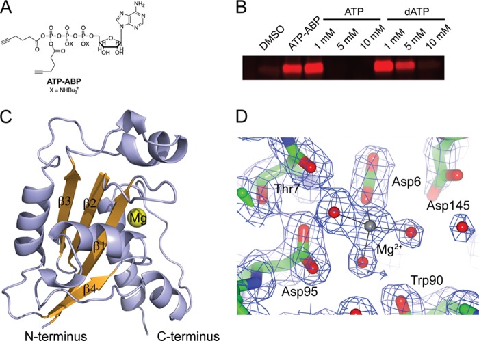
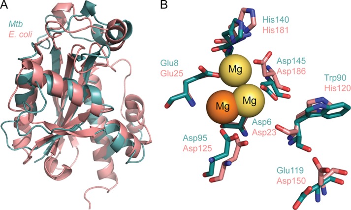
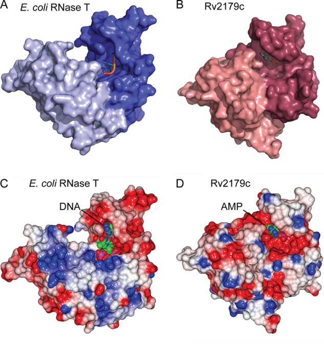
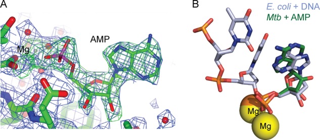
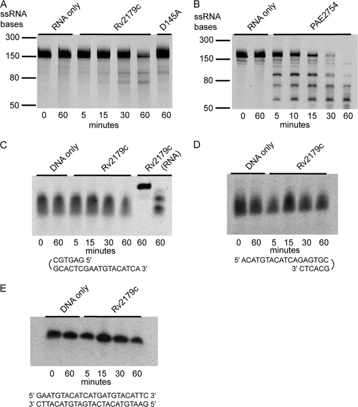
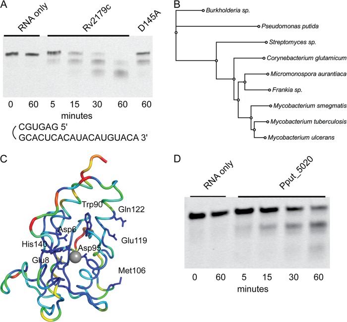
Ribonucleases (RNases) maintain the cellular RNA pool by RNA processing and degradation. In many bacteria, including the human pathogen Mycobacterium tuberculosis (Mtb), the enzymes mediating several central RNA processing functions are still unknown. Here, we identify the hypothetical Mtb protein Rv2179c as a highly divergent exoribonuclease. Although the primary sequence of Rv2179c has no detectable similarity to any known RNase, the Rv2179c crystal structure reveals an RNase fold. Active site residues are equivalent to those in the DEDD family of RNases, and Rv2179c has close structural homology to Escherichia coli RNase T. Consistent with the DEDD fold, Rv2179c has exoribonuclease activity, cleaving the 3' single-strand overhangs of duplex RNA. Functional orthologs of Rv2179c are prevalent in actinobacteria and found in bacteria as phylogenetically distant as proteobacteria. Thus, Rv2179c is the founding member of a new, large RNase family with hundreds of members across the bacterial kingdom.

Keywords: Annotation; Crystal Structure; Microbiology; Mycobacterium tuberculosis; Pseudomonas; Ribonuclease.


Figures

FIGURE 1.
FIGURE 1.
Rv2179c has an RNase fold. A, ATP-ABP activity probe used for the identification of Rv2179c. B, Rv2179c binds ATP-ABP, and binding is inhibited by ATP and dATP. C, overall structure of Rv2179c shows an RNase fold. D, 2FoFc electron density map shows active site residues and the magnesium ion contoured at 1σ. Mg2+ is coordinated by three water molecules and the carboxylate group of Asp-6.
FIGURE 2.
FIGURE 2.
Rv2179c is a structural ortholog of E. coli RNase T. A, overlay of Rv2179c and E. coli RNase T shows close similarity of topology and secondary structure elements. B, overlay of the DEDD family active site residues identifies Rv2179c as a DEDDh family member. Orange magnesium ion, Rv2179c. Yellow magnesium ions, RNase T.
FIGURE 3.
FIGURE 3.
Rv2179c forms a dimer in the crystal and in solution. A, overall orientation of the two Rv2179c protomers in the dimer. B, size exclusion chromatography showing elution of recombinant Rv2179c (molecular mass ∼20 kDa) at a molecular mass consistent with a dimer. C, dimer interface showing large complementary surfaces, together creating a 1200-Å interface. D, network of hydrogen bonds stabilizing the dimer interface.
FIGURE 4.
FIGURE 4.
The Rv2179c dimer is oriented similarly to that of RNase T. A, surface representation of the RNase T protomers. B, Rv2179c protomers. C, electrostatic surface potential of RNase T. D, Rv2179c showing a more extensive electropositive binding groove in RNase T.
FIGURE 5.
FIGURE 5.
Rv2179c binds AMP in the active site. A, omit density map for AMP in the AMP-bound structure. Both AMP and magnesium were omitted for the calculation of the maps. The σA-weighted 2FoFc map contoured at 1σ is shown in blue, the σA-weighted FoFc map contoured at 3σ is shown in green. B, Rv2179c structure in complex with AMP showing AMP binding consistent with substrate binding in E. coli RNase T.
FIGURE 6.
FIGURE 6.
Rv2179c does not have endonuclease or DNase activity. A and B, endoribonuclease assay using pentaprobes representing all possible 5-nucleotide combinations shows limited activity of Rv2179c compared with the endoribonuclease PAE2754 from Pyrobaculum aerophilum (B, Ref. 24). C, activity of Rv2179c on 3′ DNA overhangs. D and E, activity of Rv2179c on 5′ DNA overhangs (D) and on blunt-end DNA (E).
FIGURE 7.
FIGURE 7.
Rv2179c has 3′ exoribonuclease activity and establishes a new RNase family. A, exoribonuclease assay using RNA oligonucleotides with 3′ overhangs shows degradation of single strand 3′ ends. B, unrooted phylogenetic tree of nine sequences showing distribution of Rv2179c orthologs among bacterial species. C, sequence conservation in >400 Rv2179c orthologs mapped on the Rv2179c structure using ConSurf. Blue represents high conservation; red, low conservation. D, 3′ exoribonuclease activity of the distant Rv2179c ortholog Pput_5020 from P. putida with the same substrate shown in A.

References

    1. Zuo Y., Deutscher M. P. (2001) Exoribonuclease superfamilies: structural analysis and phylogenetic distribution. Nucleic Acids Res. 29, 1017–1026 - PMC - PubMed
    1. Beese L. S., Steitz T. A. (1991) Structural basis for the 3′-5′ exonuclease activity of Escherichia coli DNA polymerase I: a two-metal ion mechanism. EMBO J. 10, 25–33 - PMC - PubMed
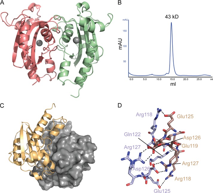
    1. Li Z., Zhan L., Deutscher M. P. (1996) Escherichia coli RNase T functions in vivo as a dimer dependent on cysteine 168. J. Biol. Chem. 271, 1133–1137 - PubMed
    1. Li Z., Deutscher M. P. (1995) The tRNA processing enzyme RNase T is essential for maturation of 5S RNA. Proc. Natl. Acad. Sci. U.S.A. 92, 6883–6886 - PMC - PubMed
    1. Li Z., Deutscher M. P. (1996) Maturation pathways for E. coli tRNA precursors: a random multienzyme process in vivo. Cell 86, 503–512 - PubMed

Publication types

MeSH terms

LinkOut - more resources